Corridon Taylor L, O'Moore Jill, Lian Yuan, Laversenne Vanessa, Noble Briana, Kamath Nikita G, Serack Fiona E, Shaikh Abdul Basit, Erickson Brian, Braun Craig, Lenz Kenney, Howard Michael, Chan Nathan, Reidenbach Andrew G, Cabin Deborah E, Vallabh Sonia M, Grindeland Andrea, Oberbeck Nina, Zhao Hien T, Minikel Eric Vallabh
Program in Brain Health, Broad Institute of MIT and Harvard, Cambridge, MA, 02142, USA.
Weissman Hood Institute, Great Falls, MT, 59405, USA.
bioRxiv. 2024 Nov 14:2024.11.12.623215. doi: 10.1101/2024.11.12.623215.
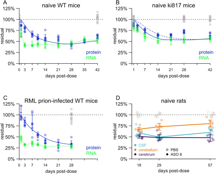
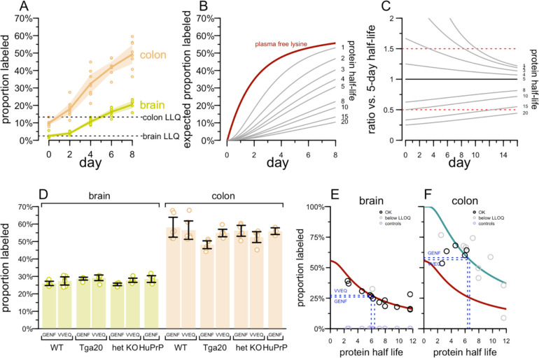
PrP lowering is effective against prion disease in animal models and is being tested clinically. Therapies in the current pipeline lower PrP production, leaving pre-existing PrP to be cleared according to its own half-life. We hypothesized that PrP's half-life may be a rate-limiting factor for the time to effect of PrP-lowering drugs, and one reason why late treatment of prion-infected mice is not as effective as early treatment. Using isotopically labeled chow with targeted mass spectrometry, as well as antisense oligonucleotide treatment followed by timed PrP measurement, we estimate a half-life of 5-6 days for PrP in the brain. PrP turnover is not affected by over- or under-expression. Mouse PrP and human PrP have similar turnover rates measured in wild-type or humanized knock-in mice. CSF PrP appears to mirror brain PrP in real time in rats. PrP is more readily quantifiable in colon than in other peripheral organs, and appears to have a shorter half-life in colon than in brain. Our data may inform the design of both preclinical and clinical studies of PrP-lowering drugs.
在动物模型中,降低朊蛋白(PrP)对朊病毒病有效,目前正在进行临床试验。现有研发流程中的疗法可降低PrP的产生,让已有的PrP根据其自身半衰期被清除。我们推测,PrP的半衰期可能是影响降低PrP药物起效时间的一个限速因素,也是朊病毒感染小鼠晚期治疗不如早期治疗有效的原因之一。通过使用同位素标记饲料结合靶向质谱分析,以及反义寡核苷酸治疗后定时测量PrP,我们估计大脑中PrP的半衰期为5 - 6天。PrP的周转不受过度表达或表达不足的影响。在野生型或人源化敲入小鼠中测量,小鼠PrP和人PrP具有相似的周转率。在大鼠中,脑脊液PrP似乎实时反映大脑PrP。与其他外周器官相比,PrP在结肠中更容易定量,并且在结肠中的半衰期似乎比在大脑中更短。我们的数据可为降低PrP药物的临床前和临床研究设计提供参考。