• 文献检索
  • 文档翻译
  • 深度研究
  • 学术资讯
  • Suppr Zotero 插件Zotero 插件
  • 邀请有礼
  • 套餐&价格
  • 历史记录
应用&插件
Suppr Zotero 插件Zotero 插件浏览器插件Mac 客户端Windows 客户端微信小程序
定价
高级版会员购买积分包购买API积分包
服务
文献检索文档翻译深度研究API 文档MCP 服务
关于我们
关于 Suppr公司介绍联系我们用户协议隐私条款
关注我们

Suppr 超能文献

核心技术专利:CN118964589B侵权必究
粤ICP备2023148730 号-1Suppr @ 2026

文献检索

告别复杂PubMed语法,用中文像聊天一样搜索,搜遍4000万医学文献。AI智能推荐,让科研检索更轻松。

立即免费搜索

文件翻译

保留排版,准确专业,支持PDF/Word/PPT等文件格式,支持 12+语言互译。

免费翻译文档

深度研究

AI帮你快速写综述,25分钟生成高质量综述,智能提取关键信息,辅助科研写作。

立即免费体验

KANK1 通过损害 Scribble 介导的 Hippo 激活促进乳腺癌的发展。

KANK1 promotes breast cancer development by compromising Scribble-mediated Hippo activation.

机构信息

Department of Molecular Medicine, Max Planck Institute of Biochemistry, Martinsried, Germany.

School of Biomedical Sciences, LKS Faculty of Medicine, The University of Hong Kong, Hong Kong SAR, China.

出版信息

Nat Commun. 2024 Nov 29;15(1):10381. doi: 10.1038/s41467-024-54645-9.

DOI:10.1038/s41467-024-54645-9
PMID:39613731
原文链接:https://pmc.ncbi.nlm.nih.gov/articles/PMC11607453/
Abstract

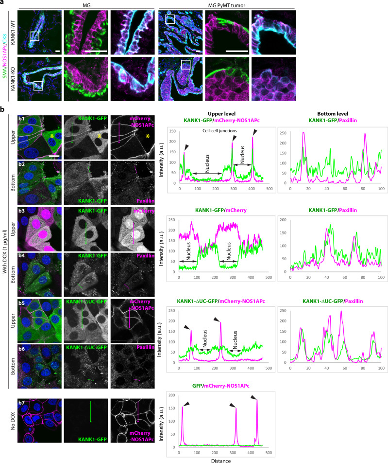
KANK1 is expressed in epithelial cells and connects focal adhesions with the adjacent cortical microtubule stabilizing complex. Although KANK1 was shown to suppress cancer cell growth in vitro, TCGA database points to high KANK1 levels associated with poor prognosis in a wide spectrum of human malignancies. Here, we address this discrepancy and report that KANK1 promotes proliferation and survival of PyMT-transformed mammary tumor cells in vivo. Mechanistically, KANK1 localizes to the basal side of basement membrane (BM)-attached transformed luminal epithelial cells. When these cells lose the contact with the BM and disassemble integrin adhesions, KANK1 is found at cell-cell junctions where it competes with the polarity and tumor suppressor Scribble for NOS1AP binding, which curbs the ability of Scribble to promote Hippo pathway activity. The consequences are stabilization and nuclear accumulation of TAZ, growth and survival of tumor cells and elevated breast cancer development.

摘要

KANK1 表达于上皮细胞,将黏着斑与相邻的皮质微管稳定复合物连接起来。尽管 KANK1 已被证明能在体外抑制癌细胞的生长,但 TCGA 数据库指出,在广泛的人类恶性肿瘤中,KANK1 水平较高与预后不良相关。在这里,我们解决了这一差异,并报告 KANK1 促进了 PyMT 转化的乳腺肿瘤细胞在体内的增殖和存活。从机制上讲,KANK1 定位于基底膜(BM)附着的转化腔上皮细胞的基底侧。当这些细胞失去与 BM 的接触并解聚整合素黏附时,KANK1 位于细胞-细胞连接处,与极性和肿瘤抑制因子 Scribble 竞争与 NOS1AP 的结合,从而抑制 Scribble 促进 Hippo 通路活性的能力。其结果是 TAZ 的稳定和核积累,肿瘤细胞的生长和存活,以及乳腺癌的发展增加。

https://cdn.ncbi.nlm.nih.gov/pmc/blobs/fb45/11607453/871f05cfcae3/41467_2024_54645_Fig10_HTML.jpg
https://cdn.ncbi.nlm.nih.gov/pmc/blobs/fb45/11607453/8f719c54fc0a/41467_2024_54645_Fig1_HTML.jpg
https://cdn.ncbi.nlm.nih.gov/pmc/blobs/fb45/11607453/1a4990641bb4/41467_2024_54645_Fig2_HTML.jpg
https://cdn.ncbi.nlm.nih.gov/pmc/blobs/fb45/11607453/da81a981f178/41467_2024_54645_Fig3_HTML.jpg
https://cdn.ncbi.nlm.nih.gov/pmc/blobs/fb45/11607453/747855ebc449/41467_2024_54645_Fig4_HTML.jpg
https://cdn.ncbi.nlm.nih.gov/pmc/blobs/fb45/11607453/8fbab8080976/41467_2024_54645_Fig5_HTML.jpg
https://cdn.ncbi.nlm.nih.gov/pmc/blobs/fb45/11607453/a3d848d74e9d/41467_2024_54645_Fig6_HTML.jpg
https://cdn.ncbi.nlm.nih.gov/pmc/blobs/fb45/11607453/d4e15cffc2ac/41467_2024_54645_Fig7_HTML.jpg
https://cdn.ncbi.nlm.nih.gov/pmc/blobs/fb45/11607453/15d5ddc3f1bf/41467_2024_54645_Fig8_HTML.jpg
https://cdn.ncbi.nlm.nih.gov/pmc/blobs/fb45/11607453/16bcc51aa15d/41467_2024_54645_Fig9_HTML.jpg
https://cdn.ncbi.nlm.nih.gov/pmc/blobs/fb45/11607453/871f05cfcae3/41467_2024_54645_Fig10_HTML.jpg

相似文献

1
KANK1 promotes breast cancer development by compromising Scribble-mediated Hippo activation.KANK1 通过损害 Scribble 介导的 Hippo 激活促进乳腺癌的发展。
Nat Commun. 2024 Nov 29;15(1):10381. doi: 10.1038/s41467-024-54645-9.
2
Loss of DLG5 promotes breast cancer malignancy by inhibiting the Hippo signaling pathway.DLG5 的缺失通过抑制 Hippo 信号通路促进乳腺癌的恶性转化。
Sci Rep. 2017 Feb 7;7:42125. doi: 10.1038/srep42125.
3
Deubiquitinating Enzyme USP9X Suppresses Tumor Growth via LATS Kinase and Core Components of the Hippo Pathway.去泛素化酶USP9X通过LATS激酶和Hippo信号通路的核心组件抑制肿瘤生长。
Cancer Res. 2017 Sep 15;77(18):4921-4933. doi: 10.1158/0008-5472.CAN-16-3413. Epub 2017 Jul 18.
4
The Hippo component YAP localizes in the nucleus of human papilloma virus positive oropharyngeal squamous cell carcinoma.河马信号通路组件YAP定位于人乳头瘤病毒阳性口咽鳞状细胞癌的细胞核中。
J Otolaryngol Head Neck Surg. 2017 Feb 22;46(1):15. doi: 10.1186/s40463-017-0187-1.
5
Blockade of a novel MAP4K4-LATS2-SASH1-YAP1 cascade inhibits tumorigenesis and metastasis in luminal breast cancer.阻断新型 MAP4K4-LATS2-SASH1-YAP1 级联反应抑制腔面型乳腺癌的肿瘤发生和转移。
J Biol Chem. 2024 Jun;300(6):107309. doi: 10.1016/j.jbc.2024.107309. Epub 2024 Apr 22.
6
Hippo pathway effectors YAP, TAZ and TEAD are associated with EMT master regulators ZEB, Snail and with aggressive phenotype in phyllodes breast tumors.Hippo 通路效应物 YAP、TAZ 和 TEAD 与 EMT 主调控因子 ZEB、Snail 以及叶状乳腺肿瘤中的侵袭表型相关。
Pathol Res Pract. 2024 Oct;262:155551. doi: 10.1016/j.prp.2024.155551. Epub 2024 Aug 15.
7
The Hippo pathway terminal effector TAZ/WWTR1 mediates oxaliplatin sensitivity in p53 proficient colon cancer cells. Hippo 通路终末效应物 TAZ/WWTR1 介导 p53 功能正常的结肠癌细胞对奥沙利铂的敏感性。
BMC Cancer. 2024 May 14;24(1):587. doi: 10.1186/s12885-024-12316-4.
8
Hippo component YAP promotes focal adhesion and tumour aggressiveness via transcriptionally activating THBS1/FAK signalling in breast cancer.Hippo 通路组件 YAP 通过转录激活 THBS1/FAK 信号促进乳腺癌中的黏附斑形成和肿瘤侵袭性。
J Exp Clin Cancer Res. 2018 Jul 28;37(1):175. doi: 10.1186/s13046-018-0850-z.
9
SnoN Antagonizes the Hippo Kinase Complex to Promote TAZ Signaling during Breast Carcinogenesis.SnoN在乳腺癌发生过程中拮抗Hippo激酶复合物以促进TAZ信号传导。
Dev Cell. 2016 Jun 6;37(5):399-412. doi: 10.1016/j.devcel.2016.05.002. Epub 2016 May 26.
10
Hippo pathway inactivation through subcellular localization of NF2/merlin in outer cells of mouse embryos.通过 NF2/merlin 在小鼠胚胎外细胞中的亚细胞定位来抑制 Hippo 通路。
Development. 2024 Jul 15;151(14). doi: 10.1242/dev.202639. Epub 2024 Jul 30.

引用本文的文献

1
Posttranslational modifications of YAP/TAZ: molecular mechanisms and therapeutic opportunities.YAP/TAZ的翻译后修饰:分子机制与治疗机遇
Cell Mol Biol Lett. 2025 Jul 17;30(1):83. doi: 10.1186/s11658-025-00760-4.

本文引用的文献

1
Discovery and ranking of the most robust prognostic biomarkers in serous ovarian cancer.发现和评估浆液性卵巢癌中最稳健的预后生物标志物。
Geroscience. 2023 Jun;45(3):1889-1898. doi: 10.1007/s11357-023-00742-4. Epub 2023 Mar 1.
2
YAP/TAZ as master regulators in cancer: modulation, function and therapeutic approaches.YAP/TAZ 作为癌症的主要调控因子:调节、功能和治疗方法。
Nat Cancer. 2023 Jan;4(1):9-26. doi: 10.1038/s43018-022-00473-z. Epub 2022 Dec 23.
3
KANK proteins.KANK蛋白
Curr Biol. 2022 Oct 10;32(19):R990-R992. doi: 10.1016/j.cub.2022.08.073.
4
Molecular analysis of TCGA breast cancer histologic types.癌症基因组图谱(TCGA)乳腺癌组织学类型的分子分析。
Cell Genom. 2021 Dec 8;1(3). doi: 10.1016/j.xgen.2021.100067.
5
Mechanoregulation of YAP and TAZ in Cellular Homeostasis and Disease Progression.YAP和TAZ在细胞稳态及疾病进展中的机械调节作用
Front Cell Dev Biol. 2021 May 24;9:673599. doi: 10.3389/fcell.2021.673599. eCollection 2021.
6
Recessive variants impair actin remodeling and cause glomerulopathy in humans and mice.隐性变异会损害肌动蛋白重塑,并导致人类和小鼠的肾小球病。
Sci Adv. 2021 Jan 1;7(1). doi: 10.1126/sciadv.abe1386. Print 2021 Jan.
7
KANK family proteins in cancer.KANK 家族蛋白与癌症。
Int J Biochem Cell Biol. 2021 Feb;131:105903. doi: 10.1016/j.biocel.2020.105903. Epub 2020 Dec 10.
8
Tissue distribution and subcellular localization of the family of Kidney Ankyrin Repeat Domain (KANK) proteins.家族性肾锚蛋白重复结构域(KANK)蛋白的组织分布与亚细胞定位。
Exp Cell Res. 2021 Jan 1;398(1):112391. doi: 10.1016/j.yexcr.2020.112391. Epub 2020 Nov 28.
9
The Scribble family in cancer: twentieth anniversary.Scribble 家族与癌症:二十周年纪念
Oncogene. 2020 Nov;39(47):7019-7033. doi: 10.1038/s41388-020-01478-7. Epub 2020 Sep 30.
10
Visualizing and interpreting cancer genomics data via the Xena platform.通过Xena平台可视化和解读癌症基因组学数据。
Nat Biotechnol. 2020 Jun;38(6):675-678. doi: 10.1038/s41587-020-0546-8.