Aukema Robert J, Petrie Gavin N, Baglot Samantha L, Gilpin Nicholas W, Hill Matthew N
Neuroscience Graduate Program, University of Calgary, Calgary, AB, T2N 4N1, Canada.
Hotchkiss Brain Institute, Cumming School of Medicine, University of Calgary, Calgary, AB, T2N 4N1, Canada.
Neurobiol Stress. 2024 Nov 15;33:100694. doi: 10.1016/j.ynstr.2024.100694. eCollection 2024 Nov.
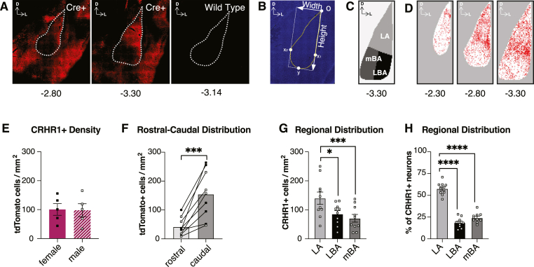
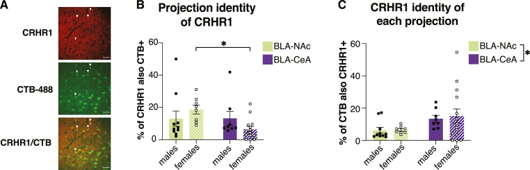
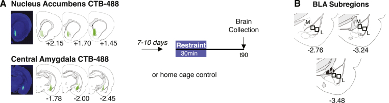
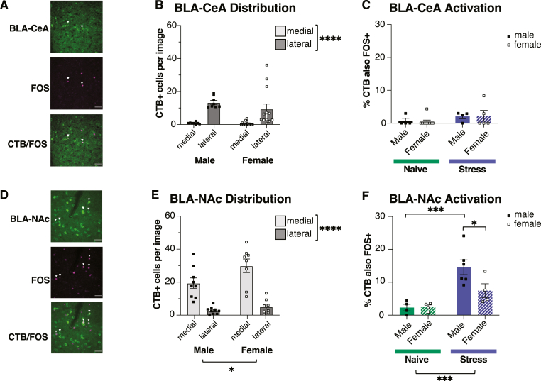
Although the basolateral amygdala (BLA) and corticotropin releasing hormone receptor type I (CRHR1) signaling are both central to the stress response, the spatial and circuit-specific distribution of CRHR1 have not been identified in the BLA at a high resolution. We used transgenic male and female CRHR1-Cre-tdTomato rats to topographically map the distribution of BLA neurons and identify whether they are activated by acute stress. Additionally, we used the BLA circuits projecting to the central amygdala (CeA) and nucleus accumbens (NAc) as a model to test circuit-specific expression of CRHR1 in the BLA. We established several key findings. First, CRHR1 had the strongest expression in the lateral amygdala and in caudal portions of the BLA. Second, acute restraint stress increased FOS expression of CRHR1 neurons, and stress-induced activation was particularly strong in medial subregions of the BLA. Third, stress significantly increased FOS expression on BLA-NAc, but not BLA-CeA projectors, and BLA-NAc activation was more robust in males than females. Finally, CRHR1 was expressed on a subset of BLA-CeA and BLA-NAc projection neurons. Collectively, this expands our understanding of BLA molecular- and circuit-specific activation patterns following acute stress.
虽然基底外侧杏仁核(BLA)和促肾上腺皮质激素释放激素受体I型(CRHR1)信号传导在应激反应中都起着核心作用,但尚未在高分辨率下确定BLA中CRHR1的空间和回路特异性分布。我们使用转基因雄性和雌性CRHR1-Cre-tdTomato大鼠对BLA神经元的分布进行拓扑映射,并确定它们是否被急性应激激活。此外,我们以投射到中央杏仁核(CeA)和伏隔核(NAc)的BLA回路为模型,测试BLA中CRHR1的回路特异性表达。我们得出了几个关键发现。第一,CRHR1在杏仁核外侧和BLA的尾部表达最强。第二,急性束缚应激增加了CRHR1神经元的FOS表达,并且应激诱导的激活在BLA的内侧亚区域特别强烈。第三,应激显著增加了BLA-NAc投射神经元上的FOS表达,但未增加BLA-CeA投射神经元上的FOS表达,并且BLA-NAc的激活在雄性中比雌性中更强。最后,CRHR1在BLA-CeA和BLA-NAc投射神经元的一个子集中表达。总的来说,这扩展了我们对急性应激后BLA分子和回路特异性激活模式的理解。