Saglam-Metiner Pelin, Yanasik Sena, Odabasi Yusuf Caglar, Modamio Jennifer, Negwer Moritz, Biray-Avci Cigir, Guler Ayse, Erturk Ali, Yildirim Ender, Yesil-Celiktas Ozlem
Department of Bioengineering, Faculty of Engineering, Ege University, Izmir, Türkiye.
Institute for Tissue Engineering and Regenerative Medicine (iTERM), Helmholtz Zentrum München, Neuherberg, Germany.
Commun Biol. 2024 Dec 5;7(1):1627. doi: 10.1038/s42003-024-07313-z.
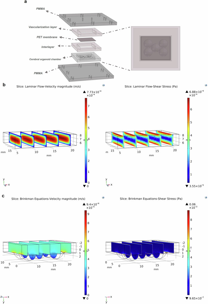
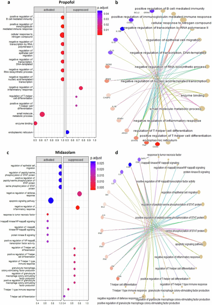
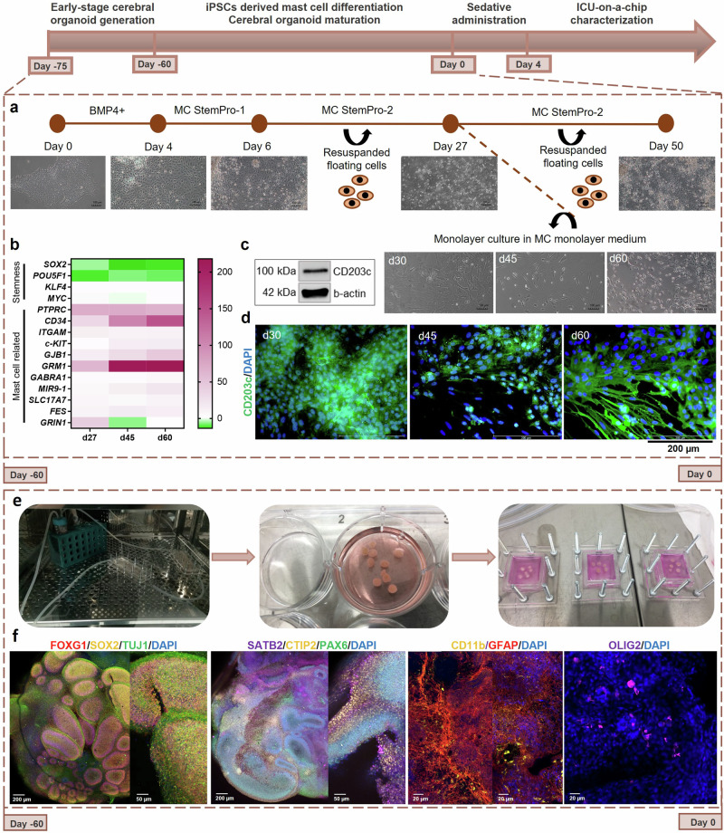
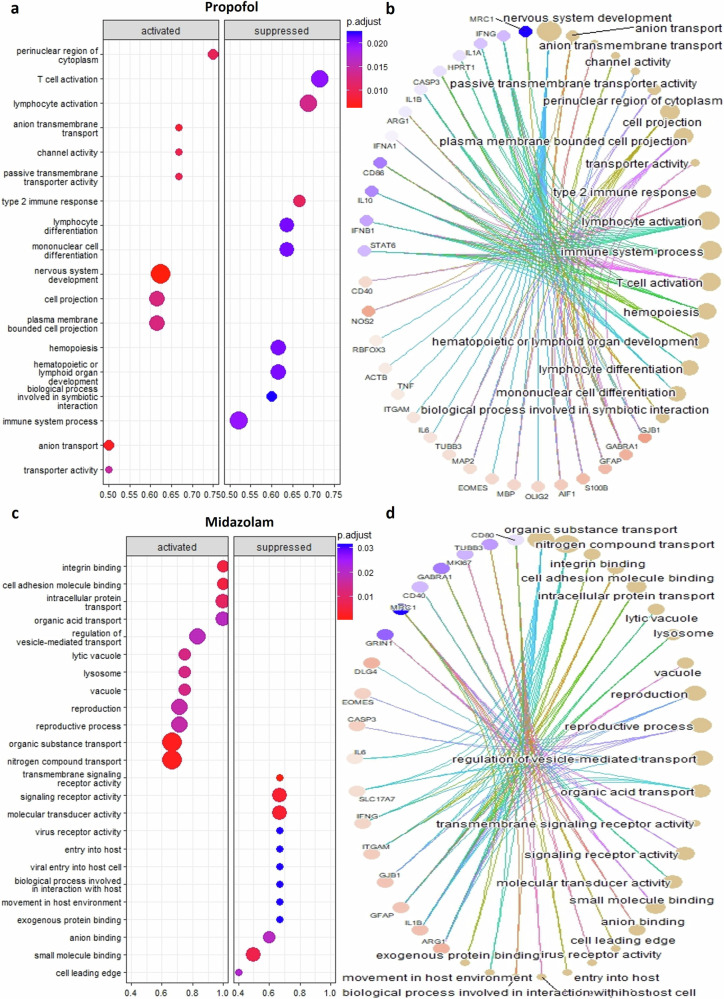
Propofol and midazolam are the current standard of care for prolonged sedation in Intensive Care Units (ICUs). However, the effects and mechanism of these sedatives in brain tissue are unclear. Herein, the development of an ICU patient-on-a-chip platform to elucidate those effects is reported. The humanized neural tissue compartment combines mast cells differentiated from human induced pluripotent stem cells (hiPSCs) with cerebral organoids in a three-dimensional (3D) matrix, which is covered with a membrane populated with human cerebral microvascular endothelial cells (hCMEC/D3) that separates the tissue chamber from the vascular lumen, where sedatives were infused for four days to evaluate neurotoxicity and cell-mediated immune responses. Subsequent to propofol administration, gene expressions of CD40 and TNF-α in mast cells, AIF1 in microglia and GFAP/S100B/OLIG2/MBP in macroglia were elevated, as well as NOS2, CD80, CD40, CD68, IL6 and TNF-α mediated proinflammation is noted in cerebral organoids, which resulted in higher expressions of GJB1, GABA-A and NMDAR1 in the tissue construct of the platform. Besides, midazolam administration stimulated expression of CD40 and CD203c+ reactivated mast cell proliferation and compromised BBB permeability and decreased TEER values with higher barrier disruption, whereas increased populations of CD11b+ microglia, higher expressions of GFAP/DLG4/GJB1 and GABA-A-/NMDAR1- identities, as well as glutamate related neurotoxicity and IL1B, IFNG, IFNA1, IL6 genes mediated proinflammation, resulting in increased apoptotic zones are observed in cerebral organoids. These results suggest that different sedatives cause variations in cell type activation that modulate different pathways related to neuroinflammation and neurotoxicity in the ICU patient-on-chip platform.
丙泊酚和咪达唑仑是目前重症监护病房(ICU)长期镇静的标准治疗药物。然而,这些镇静剂在脑组织中的作用和机制尚不清楚。在此,报告了一种用于阐明这些作用的ICU芯片上患者平台的开发。人源化神经组织隔室将从人诱导多能干细胞(hiPSC)分化而来的肥大细胞与三维(3D)基质中的脑类器官相结合,该基质覆盖有一层由人脑血管内皮细胞(hCMEC/D3)构成的膜,该膜将组织腔与血管腔分隔开,在血管腔中注入镇静剂四天以评估神经毒性和细胞介导的免疫反应。给予丙泊酚后,肥大细胞中CD40和TNF-α的基因表达、小胶质细胞中AIF1的基因表达以及大胶质细胞中GFAP/S100B/OLIG2/MBP的基因表达均升高,并且在脑类器官中观察到NOS2、CD80、CD40、CD68、IL6和TNF-α介导的促炎反应,这导致平台组织构建体中GJB1、GABA-A和NMDAR1的表达升高。此外,给予咪达唑仑刺激了CD40和CD203c+再活化肥大细胞增殖的表达,损害了血脑屏障通透性并降低了跨上皮电阻值,同时屏障破坏增加,而CD11b+小胶质细胞数量增加、GFAP/DLG4/GJB1和GABA-A-/NMDAR1-身份的表达升高,以及谷氨酸相关神经毒性和IL1B、IFNG、IFNA1、IL6基因介导的促炎反应,导致在脑类器官中观察到凋亡区域增加。这些结果表明,不同的镇静剂会导致细胞类型激活的差异,从而调节ICU芯片上患者平台中与神经炎症和神经毒性相关的不同途径。