Tan Jerome, Chen Jiahui, Roxby Daniel, Chooi Wai Hon, Nguyen Tan Dai, Ng Shi Yan, Han Jongyoon, Chew Sing Yian
School of Chemistry, Chemical Engineering and Biotechnology, Nanyang Technological University, Singapore, Singapore.
HealthTech @ NTU, Interdisciplinary Graduate Programme, Nanyang Technological University, Singapore, Singapore.
Stem Cell Res Ther. 2024 Dec 5;15(1):465. doi: 10.1186/s13287-024-04070-y.
The emergence of induced pluripotent stem cells (iPSCs) offers a promising approach for replacing damaged neurons and glial cells, particularly in spinal cord injuries (SCI). Despite its merits, iPSC differentiation into spinal cord progenitor cells (SCPCs) is variable, necessitating reliable assessment of differentiation and validation of cell quality and safety. Phenotyping is often performed via label-based methods including immunofluorescent staining or flow cytometry analysis. These approaches are often expensive, laborious, time-consuming, destructive, and severely limits their use in large scale cell therapy manufacturing settings. On the other hand, cellular biophysical properties have demonstrated a strong correlation to cell state, quality and functionality and can be measured with ingenious label-free technologies in a rapid and non-destructive manner.
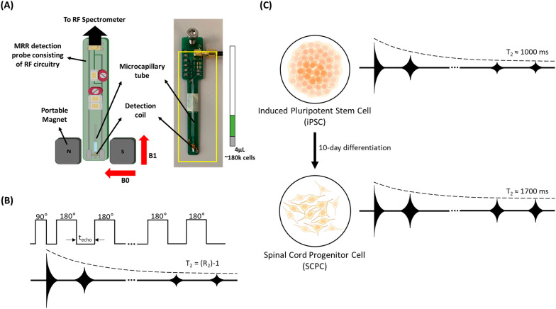
In this study, we report the use of Magnetic Resonance Relaxometry (MRR), a rapid and label-free method that indicates iron levels based on its readout (T). Briefly, we differentiated human iPSCs into SCPCs and compared key iPSC and SCPC cellular markers to their intracellular iron content (Fe) at different stages of the differentiation process.
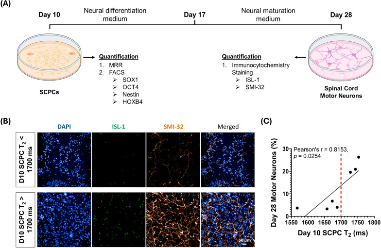
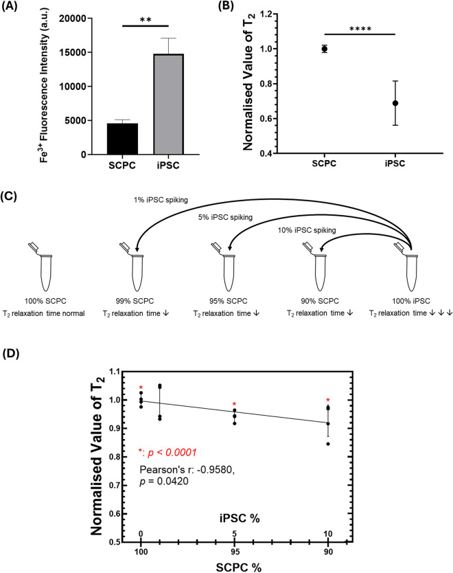
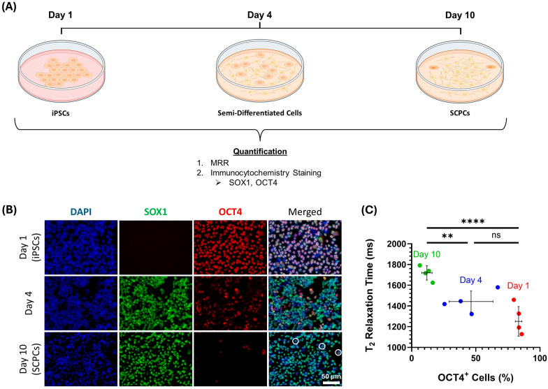
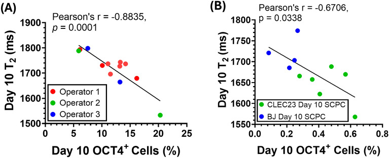
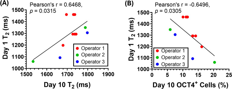
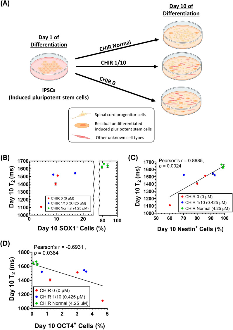
With MRR, we found that intracellular iron of iPSCs and SCPCs were distinctively different allowing us to accurately reflect varying levels of residual undifferentiated iPSCs (i.e., OCT4 cells) in any given population of SCPCs. MRR was also able to predict Day 10 SCPC OCT4 levels from Day 1 undifferentiated iPSC T values and identified poorly differentiated SCPCs with lower T, indicative of lower neural progenitor (SOX1) and stem cell (Nestin) marker expression levels. Lastly, MRR was able to provide predictive indications for the extent of differentiation to Day 28 spinal cord motor neurons (ISL-1/SMI-32) based on the T values of Day 10 SCPCs.
MRR measurements of iPSCs and SCPCs has clearly indicated its capabilities to identify and quantify key phenotypes of iPSCs and SCPCs for end-point validation of safety and quality parameters. Thus, our technology provides a rapid label-free method to determine critical quality attributes in iPSC-derived progenies and is ideally suited as a quality control tool in cell therapy manufacturing.
诱导多能干细胞(iPSC)的出现为替代受损神经元和神经胶质细胞提供了一种很有前景的方法,尤其是在脊髓损伤(SCI)方面。尽管有其优点,但iPSC向脊髓祖细胞(SCPC)的分化是可变的,因此需要可靠地评估分化情况并验证细胞质量和安全性。表型分析通常通过基于标记的方法进行,包括免疫荧光染色或流式细胞术分析。这些方法通常成本高昂、费力、耗时、具有破坏性,并且严重限制了它们在大规模细胞治疗生产环境中的应用。另一方面,细胞生物物理特性已证明与细胞状态、质量和功能密切相关,并且可以通过巧妙的无标记技术以快速且无损的方式进行测量。
在本研究中,我们报告了磁共振弛豫测量法(MRR)的应用,这是一种基于其读数(T)指示铁水平的快速无标记方法。简而言之,我们将人iPSC分化为SCPC,并在分化过程的不同阶段将关键的iPSC和SCPC细胞标志物与其细胞内铁含量(Fe)进行比较。
通过MRR,我们发现iPSC和SCPC的细胞内铁明显不同,这使我们能够准确反映任何给定SCPC群体中残留未分化iPSC(即OCT4细胞)的不同水平。MRR还能够根据第1天未分化iPSC的T值预测第10天SCPC的OCT4水平,并识别出T值较低的分化不良的SCPC,这表明神经祖细胞(SOX1)和干细胞(巢蛋白)标志物表达水平较低。最后,MRR能够根据第10天SCPC的T值为第28天脊髓运动神经元(ISL-1/SMI-32)的分化程度提供预测指标。
对iPSC和SCPC进行MRR测量清楚地表明了其识别和量化iPSC和SCPC关键表型以进行安全性和质量参数终点验证的能力。因此,我们的技术提供了一种快速的无标记方法来确定iPSC衍生后代的关键质量属性,非常适合作为细胞治疗生产中的质量控制工具。