Ardon Monica, Nguyen Lily, Chen Rui, Rogers Jeffrey, Stout Tim, Thomasy Sara, Moshiri Ala
Department of Ophthalmology & Vision Science, School of Medicine, University of California Davis, Sacramento, California, United States.
Human Genome Sequencing Center and Department of Molecular and Human Genetics, Baylor College of Medicine, Houston, Texas, United States.
Invest Ophthalmol Vis Sci. 2024 Dec 2;65(14):16. doi: 10.1167/iovs.65.14.16.
The California National Primate Research Center contains a colony of rhesus macaques with a homozygous missense mutation in PDE6C (R565Q) which causes a cone disorder similar to PDE6C achromatopsia in humans. The purposes of this study are to characterize the phenotype in PDE6C macaques in detail to determine the onset of the cone phenotype, the degree to which the phenotype progresses, if heterozygote animals have an intermediate phenotype, and if rod photoreceptor function declines over time.
We analyzed spectral-domain optical coherence tomography (SD-OCT), fundus autofluorescence (FAF), and electroretinography (ERG) data from 102 eyes of 51 macaques (aged 0.25 to 16 years). Measurements of retinal layers as well as cone and rod function over time were quantitatively compared.
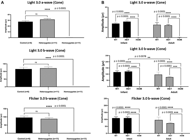
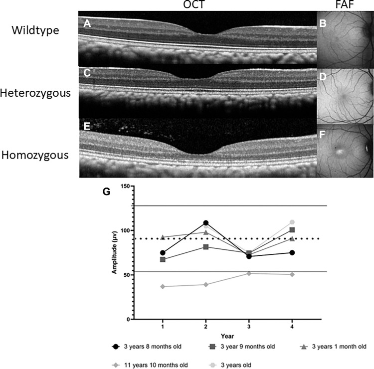
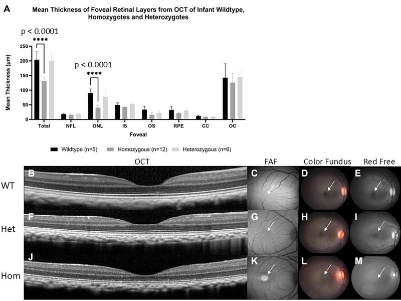
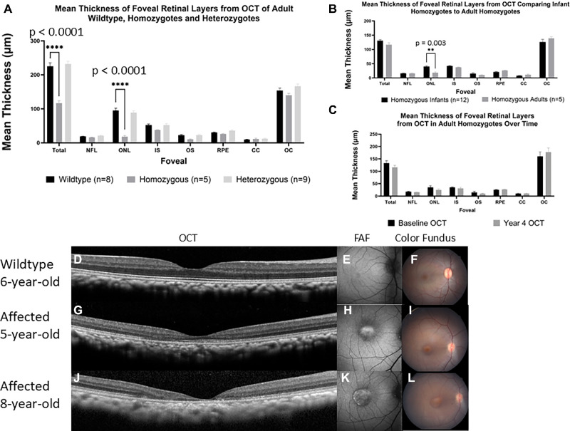
Homozygotes as young as 3 months postnatal showed absent cone responses on electroretinogram. Infant homozygotes had reduced foveal outer nuclear layer (ONL) thickness compared with wildtype infants (P < 0.0001). Over 4 years of study, no consistent changes in retinal layer thicknesses were found within 5 adult homozygotes. However, comparisons between infants and adults revealed reductions in foveal ONL thickness suggesting that cone cells slowly degenerate as homozygotes age. The oldest homozygote (11 years) had reduced rod responses. Heterozygotes could not be distinguished from wildtypes in any parameters.
These data suggest that, like humans, macaque PDE6C heterozygotes are normal, and homozygote primates have absent cone function and reduced foveal ONL thickness from infancy. Cone photoreceptors probably degenerate over time and macular atrophy can occur. Rod photoreceptor function may wane in late stages.
加利福尼亚国家灵长类研究中心饲养了一群恒河猴,其PDE6C基因存在纯合错义突变(R565Q),导致一种类似于人类PDE6C色盲症的视锥细胞疾病。本研究的目的是详细描述PDE6C基因猕猴的表型,以确定视锥细胞表型的发病时间、表型进展程度、杂合子动物是否具有中间表型,以及视杆细胞光感受器功能是否随时间下降。
我们分析了51只猕猴(年龄0.25至16岁)102只眼睛的光谱域光学相干断层扫描(SD-OCT)、眼底自发荧光(FAF)和视网膜电图(ERG)数据。对视网膜各层以及视锥和视杆细胞功能随时间的测量进行了定量比较。
出生后3个月大的纯合子在视网膜电图上显示视锥细胞反应缺失。与野生型婴儿相比,纯合子婴儿的中央凹外核层(ONL)厚度降低(P < 0.0001)。在4年的研究中,5只成年纯合子的视网膜层厚度没有发现一致的变化。然而,婴儿和成年人之间的比较显示中央凹ONL厚度降低,这表明随着纯合子年龄的增长,视锥细胞会缓慢退化。最年长的纯合子(11岁)视杆细胞反应降低。在任何参数上,杂合子与野生型都无法区分。
这些数据表明,与人类一样,猕猴PDE6C杂合子是正常的,纯合子灵长类动物从婴儿期就缺乏视锥细胞功能,中央凹ONL厚度降低。视锥光感受器可能会随着时间退化,并且可能会发生黄斑萎缩。视杆光感受器功能可能在后期减弱。