Satake Tomoyuki, Morizane Chigusa, Okada Mao, Nishioka Mariko, Hiraoka Nobuyoshi, Nara Satoshi, Kakegawa Tomoya, Kobayashi Maki, Koyama Kumiko, Esaki Minoru, Okusaka Takuji
Department of Hepatobiliary and Pancreatic Oncology, National Cancer Center Hospital East, Kashiwa, Japan.
Department of Hepatobiliary and Pancreatic Oncology, National Cancer Center Hospital, Tokyo, Japan.
Cancer Med. 2024 Dec;13(23):e70474. doi: 10.1002/cam4.70474.
Although activation of human epidermal growth factor receptor 3 (HER3) is linked to resistance to targeted therapies in several cancer types, the HER3 expression profile during pancreatic cancer treatment remains unknown.
We evaluated the HER3 expression status after chemotherapy for pancreatic cancer and its association with clinicopathological features and clinical outcomes.
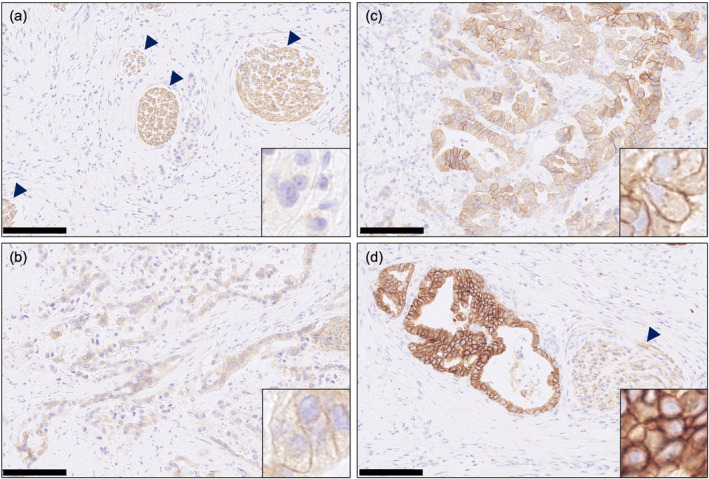
MATERIALS & METHODS: We included patients with pancreatic cancer who underwent chemotherapy and whose post-treatment archival tissue specimens were collected. HER3 expression was retrospectively assessed by immunohistochemistry scoring (0, 1+, 2+, and 3+) of the membranous staining intensity.
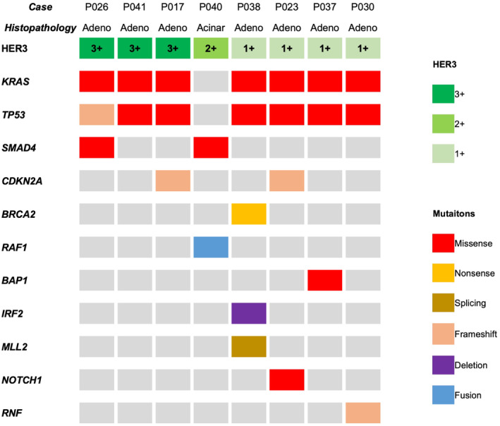
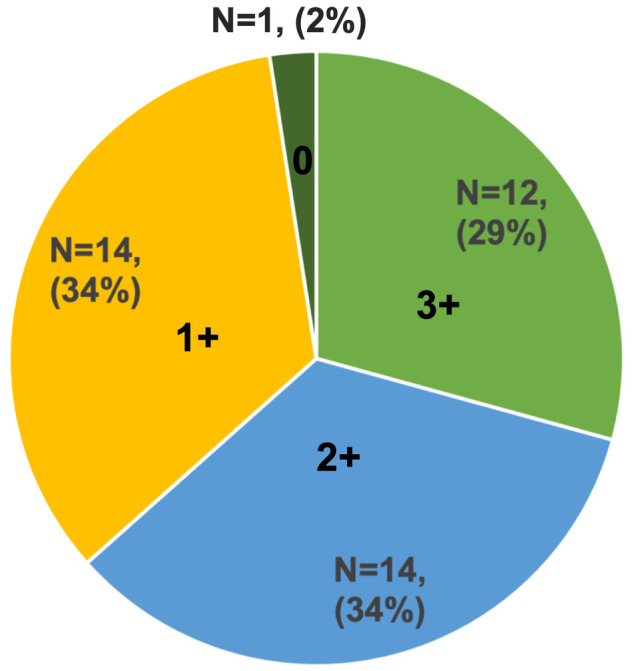
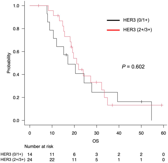
HER3 expression after chemotherapy was evaluated in 41 patients, with matched-pair analysis in five patients before and after chemotherapy. HER3 expression was observed in most of the patients after chemotherapy, demonstrating IHC scores of ≥ 1+ and ≥ 2+ in 40 (98%) and 26 (63%) of 41 patients, respectively. Of the 38 patients with adenocarcinoma, the median overall survival in the HER3 (2+/3+) and HER3 (0/1+) groups was 21.0 and 17.1 months, respectively. The comparison of HER3 expression before and after chemotherapy performed in five cases revealed that scores changed from 2+/3+ to 0/1+ in one case, 0/1+ to 2+/3+ in another case, and remained at 2+/3+ in three cases. Cancer genome profiling tests in eight cases found no HER3 amplification or mutation, and seven of these cases had adenocarcinomas with KRAS and TP53 mutations.
A high prevalence of HER3 expression was observed in pancreatic cancer patients after chemotherapy. Our findings indicate that HER3 is a potential therapeutic target for pancreatic cancer, deserving further clinical investigation.
尽管在多种癌症类型中,人表皮生长因子受体3(HER3)的激活与对靶向治疗的耐药性相关,但胰腺癌治疗期间HER3的表达谱仍不清楚。
我们评估了胰腺癌化疗后HER3的表达状态及其与临床病理特征和临床结局的关联。
我们纳入了接受化疗且收集了治疗后存档组织标本的胰腺癌患者。通过对膜染色强度进行免疫组织化学评分(0、1+、2+和3+)对HER3表达进行回顾性评估。
对41例患者化疗后的HER3表达进行了评估,其中5例患者在化疗前后进行了配对分析。化疗后大多数患者观察到HER3表达,41例患者中分别有40例(98%)和26例(63%)的免疫组化评分为≥1+和≥2+。在38例腺癌患者中,HER3(2+/3+)组和HER3(0/1+)组的中位总生存期分别为21.0个月和17.1个月。对5例患者化疗前后的HER3表达进行比较,发现1例患者评分从2+/3+变为0/1+,另1例从0/1+变为2+/3+,3例保持在2+/3+。8例患者的癌症基因组分析检测未发现HER3扩增或突变,其中7例为伴有KRAS和TP53突变的腺癌。
化疗后的胰腺癌患者中观察到HER3高表达。我们的研究结果表明,HER3是胰腺癌的一个潜在治疗靶点,值得进一步临床研究。