Zhang Donglin, Sánchez-Espino Luis Fernando, Ivars Marta, Pope Elena, Nopper Amy J, Arkin Lisa M, Tollefson Megha M, Lavarino Cinzia E, Muldowney Maya, Olaciregui Nagore Gené, Paco Sonia, Drolet Beth A, Baselga Eulàlia
Department of Dermatology, School of Medicine and Public Health, University of Wisconsin, Madison, Wisconsin, USA.
Dermatology Department, Stollery Children's Hospital, Edmonton, Alberta, Canada.
Pediatr Dermatol. 2025 May-Jun;42(3):475-480. doi: 10.1111/pde.15802. Epub 2024 Dec 9.
Many vascular anomalies harbor postzygotic somatic variants in GNAQ and GNA11; however, the phenotype of specific G-protein variants has not been well described. We report the clinical characteristics of 17 patients with a GNA11 R183C variant.
This case series is derived from a multinational cohort of vascular anomaly patients whose pathogenic mutations were identified using high-depth next generation sequencing. Data include vascular anomaly features, imaging reports, and extracutaneous manifestations of the GNA11 R183C variant.
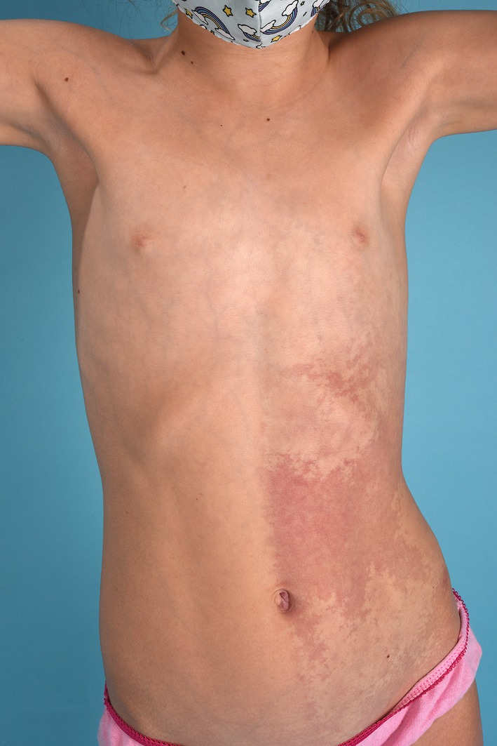
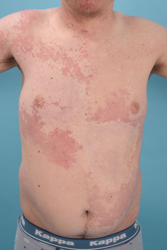
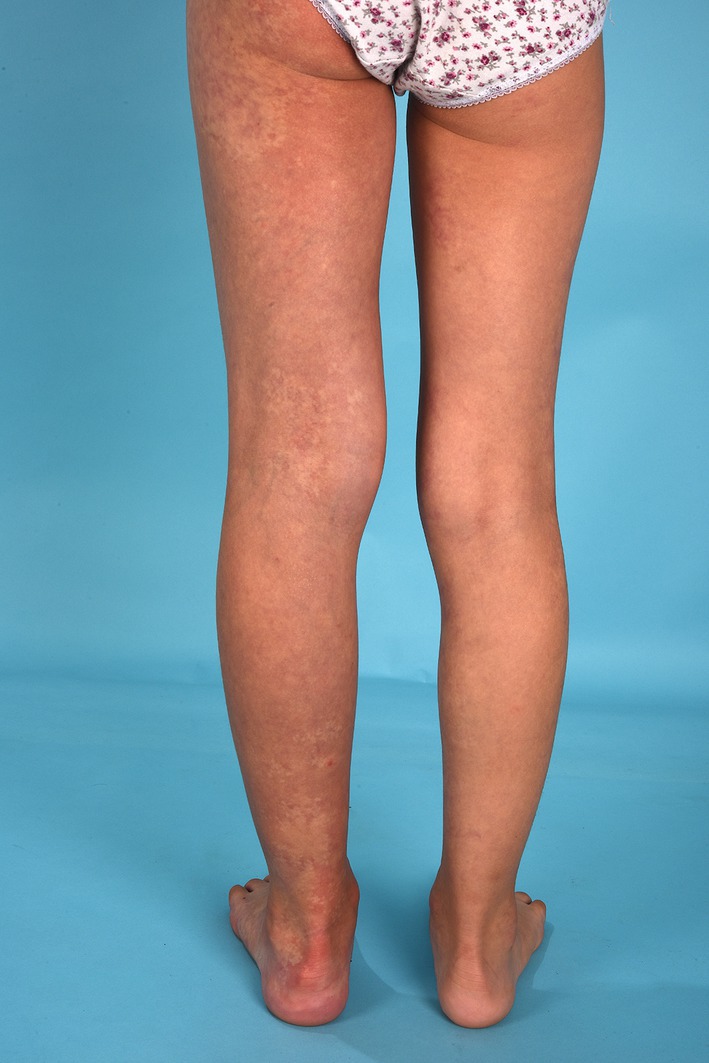
We identified 17 subjects (median age 18 years [range 6-67]) with somatic GNA11 R183C variant. All patients had vascular lesions of the skin that presented as pink-to-red in children and deeper red in adults. Most lesions were large, poorly demarcated, and reticulated patches that were often bilaterally distributed. Nevus anemicus was observed in 53% (N = 9) and dermal melanocytosis in 13.3% (N = 2) of individuals. 82% (N = 14) of patients had limb growth discrepancies, and 1 patient had marked thoracic hypoplasia. 47% (N = 8) of patients had facial involvement, and 41% (N = 7) had forehead involvement. One patient experienced seizures due to right hemispheric leptomeningeal angiomatosis consistent with Sturge-Weber syndrome. Other findings included glaucoma (29%, N = 5) and psychomotor delay (29%, N = 5).
These findings contribute to our understanding of the clinical spectrum of GNA11 R183C capillary malformations (CMs); patients characteristically present with extensive, bilateral, poorly demarcated, pink-to-red CMs associated with nevus anemicus. Glaucoma and growth discrepancies (overgrowth or undergrowth) are common. Leptomeningeal angiomatosis and developmental delay can occur, appearing potentially less prevalent and severe than GNAQ-associated disease.
许多血管异常都存在GNAQ和GNA11基因的合子后体细胞变异;然而,特定G蛋白变异的表型尚未得到充分描述。我们报告了17例携带GNA11 R183C变异患者的临床特征。
该病例系列来自一个跨国血管异常患者队列,其致病突变通过高深度下一代测序确定。数据包括血管异常特征、影像学报告以及GNA11 R183C变异的皮肤外表现。
我们确定了17名携带体细胞GNA11 R183C变异的受试者(中位年龄18岁[范围6 - 67岁])。所有患者皮肤均有血管病变,儿童表现为粉红色至红色,成人表现为深红色。大多数病变为大的、边界不清的网状斑块,常双侧分布。53%(N = 9)的个体出现贫血痣,13.3%(N = 2)的个体出现真皮黑素细胞增多症。82%(N = 14)的患者有肢体生长差异,1例患者有明显的胸廓发育不全。47%(N = 8)的患者面部受累,41%(N = 7)的患者前额受累。1例患者因与斯特奇 - 韦伯综合征一致的右侧半球软脑膜血管瘤病而癫痫发作。其他发现包括青光眼(29%,N = 5)和精神运动发育迟缓(29%,N = 5)。
这些发现有助于我们了解GNA11 R183C毛细血管畸形(CMs)的临床谱;患者的特征是出现广泛、双侧、边界不清、粉红色至红色的CMs,并伴有贫血痣。青光眼和生长差异(过度生长或生长不足)很常见。软脑膜血管瘤病和发育迟缓可能发生,其发生率和严重程度可能低于与GNAQ相关的疾病。