Mansoor Mahsaw, Ahmad Noor-Us-Sabah, Ahmed S Bilal, Tadros Samuel, Folk James, Abramoff Michael D
Department of Ophthalmology and Visual Sciences, University of Iowa, Iowa City, IA, USA.
Iowa City Veterans Affairs Medical Center, Iowa City, IA, USA.
Transl Vis Sci Technol. 2024 Dec 2;13(12):13. doi: 10.1167/tvst.13.12.13.
To investigate the efficacy of a novel approach using a sterile caliper for anterior chamber (AC) decompression in reducing post-intravitreal injection (IVI) intraocular pressure (IOP) spikes.
A prospective interventional case series conducted at the Iowa City Veterans Affairs Medical Center (VAMC) with Institutional Review Board approval. Patients were randomized to receive conventional IVI or IVI with sterile caliper decompression. Fifty eyes from 47 patients underwent IVI for various retinal pathologies. Subjects were randomly assigned to the intervention or control arm. Two resident physician providers performed injections, with one applying sterile caliper decompression (intervention) and the other following the standard technique (control). Baseline and postinjection IOP were measured using Tonopen (Reichert, Depew, NY).
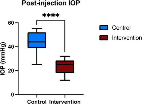
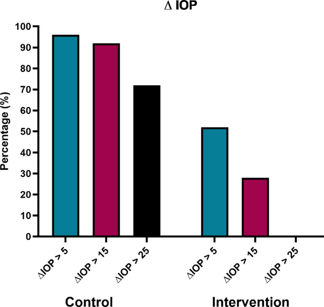
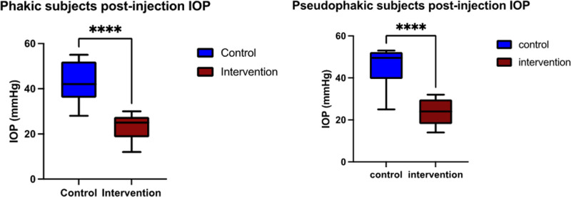
In both groups there was a significant IOP rise following IVI (P < 0.0001). There was no significant difference in baseline IOP between groups (P = 0.082), but postinjection IOP was significantly lower in the intervention group (23.52 ± 5.98 mm Hg) compared to the control group (44.08 ± 8.48 mm Hg). There were no patients with an IOP spike >25 mm Hg in the intervention arm. The technique was effective regardless of lens status.
Sterile caliper AC decompression significantly reduced post-IVI IOP spikes presenting an efficient and cost-effective alternative to previously proposed methods of IOP reduction. Further studies are warranted to validate these findings and explore broader applications in ophthalmic interventions.
The caliper decompression technique presents potential benefit in preventing short-term morbidity associated with IOP spikes after IVI and addressing long-term concerns in patients with pre-existing glaucoma.
研究一种使用无菌卡尺进行前房(AC)减压的新方法在降低玻璃体内注射(IVI)后眼压(IOP)峰值方面的疗效。
在爱荷华市退伍军人事务医疗中心(VAMC)进行的一项前瞻性干预病例系列研究,已获得机构审查委员会批准。患者被随机分为接受传统IVI或接受无菌卡尺减压的IVI。47例患者的50只眼因各种视网膜病变接受了IVI。受试者被随机分配到干预组或对照组。两名住院医师进行注射,一人应用无菌卡尺减压(干预),另一人采用标准技术(对照)。使用眼压笔(Reichert,Depew,纽约)测量基线眼压和注射后眼压。
两组在IVI后眼压均显著升高(P < 0.0001)。两组之间的基线眼压无显著差异(P = 0.082),但干预组注射后的眼压(23.52 ± 5.98 mmHg)显著低于对照组(44.08 ± 8.48 mmHg)。干预组没有眼压峰值>25 mmHg的患者。无论晶状体状态如何,该技术均有效。
无菌卡尺前房减压显著降低了IVI后的眼压峰值,是一种比先前提出的降低眼压方法更有效且具成本效益的替代方法。有必要进行进一步研究以验证这些发现,并探索其在眼科干预中的更广泛应用。
卡尺减压技术在预防IVI后眼压峰值相关的短期发病率以及解决既往存在青光眼患者的长期问题方面具有潜在益处。