• 文献检索
  • 文档翻译
  • 深度研究
  • 学术资讯
  • Suppr Zotero 插件Zotero 插件
  • 邀请有礼
  • 套餐&价格
  • 历史记录
应用&插件
Suppr Zotero 插件Zotero 插件浏览器插件Mac 客户端Windows 客户端微信小程序
定价
高级版会员购买积分包购买API积分包
服务
文献检索文档翻译深度研究API 文档MCP 服务
关于我们
关于 Suppr公司介绍联系我们用户协议隐私条款
关注我们

Suppr 超能文献

核心技术专利:CN118964589B侵权必究
粤ICP备2023148730 号-1Suppr @ 2026

文献检索

告别复杂PubMed语法,用中文像聊天一样搜索,搜遍4000万医学文献。AI智能推荐,让科研检索更轻松。

立即免费搜索

文件翻译

保留排版,准确专业,支持PDF/Word/PPT等文件格式,支持 12+语言互译。

免费翻译文档

深度研究

AI帮你快速写综述,25分钟生成高质量综述,智能提取关键信息,辅助科研写作。

立即免费体验

急性肺损伤后,募集的肺泡巨噬细胞通过产生激活素-A导致肺纤维化加重。

Exacerbation of pulmonary fibrosis following acute lung injury via activin-A production by recruited alveolar macrophages.

作者信息

Pan Ting, Feng Yinzhou, Li Yufan, Yang Yanping, Zhou Jian, Song Yuanlin

机构信息

Shanghai Key Laboratory of Lung Inflammation and Injury, Department of Pulmonary Medicine, Zhongshan Hospital, Fudan University, Shanghai, China.

Key Laboratory of Chemical Injury, Emergency and Critical Medicine of Shanghai Municipal Health Commission, Center of Emergency and Critical Medicine, Jinshan Hospital of Fudan University, Shanghai, China.

出版信息

J Thorac Dis. 2024 Nov 30;16(11):7709-7728. doi: 10.21037/jtd-24-680. Epub 2024 Nov 14.

DOI:10.21037/jtd-24-680
PMID:39678869
原文链接:https://pmc.ncbi.nlm.nih.gov/articles/PMC11635239/
Abstract

BACKGROUND

Acute respiratory distress syndrome (ARDS) is a complicated pathological cascade process of excessive pulmonary inflammation and alveolar epithelial cell apoptosis that results in respiratory dysfunction and failure. Some cases of ARDS can result in a more severe state of pulmonary fibrosis, referred to as postinjury lung fibrosis. The mortality and incidence rate of ARDS are high, particularly when it leads to continuing alveolar and interstitial fibrosis, which requires urgent treatment and appropriate management. The lipopolysaccharide (LPS)-induced acute lung injury (ALI) mouse model has been widely implemented for studying ARDS in humans. In our study, we found alterations in the alveolar macrophage (AM) profile in such a mouse model. Specifically, activin-A produced by dominantly recruited AMs (recAMs) was noted to be implicated in the process of post-injury lung fibrosis.

METHODS

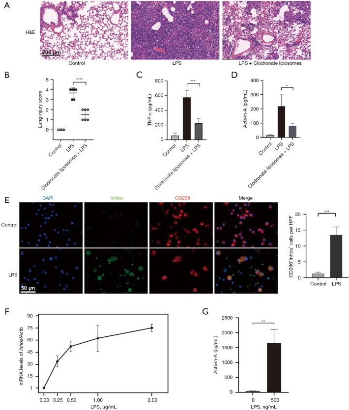
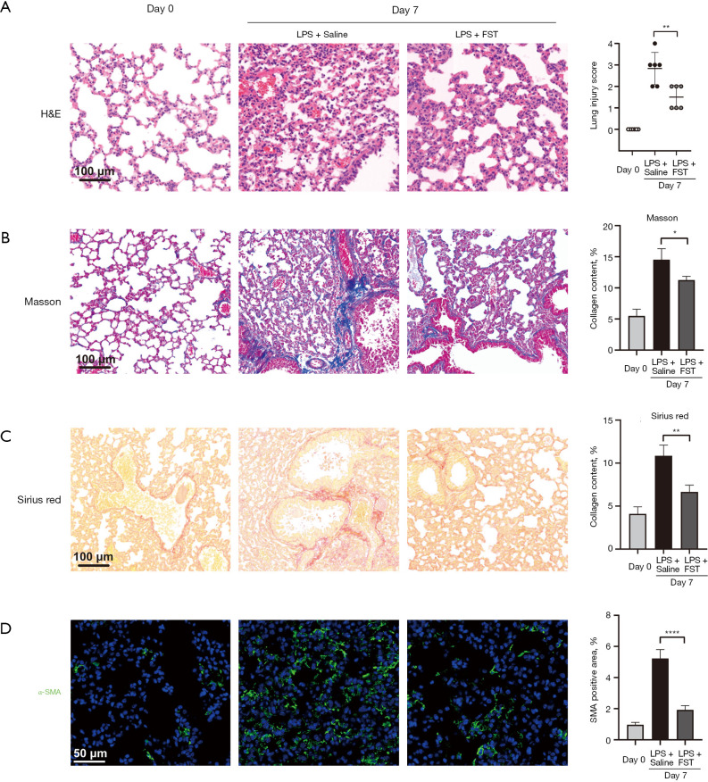
The ALI animal model in C57BL/6 mice was established via 3.5 mg/kg of LPS intratracheal administration. Single-cell RNA (scRNA) sequencing was used for detailed classification and functional characterization of lung macrophages. Through experiments, we evaluated the role that activin-A plays in post-injury lung fibrosis in an ALI mouse model using enzyme-linked immunosorbent assay (ELISA), histological staining methods, and immunofluorescence. Through experiments, we analyzed the effect of activin-A on murine lung epithelial 12 (MLE-12) cells and bone marrow-derived macrophages (BMDMs) using Western blotting (WB), quantitative real-time polymerase chain reaction, RNA sequencing, and immunofluorescence.

RESULTS

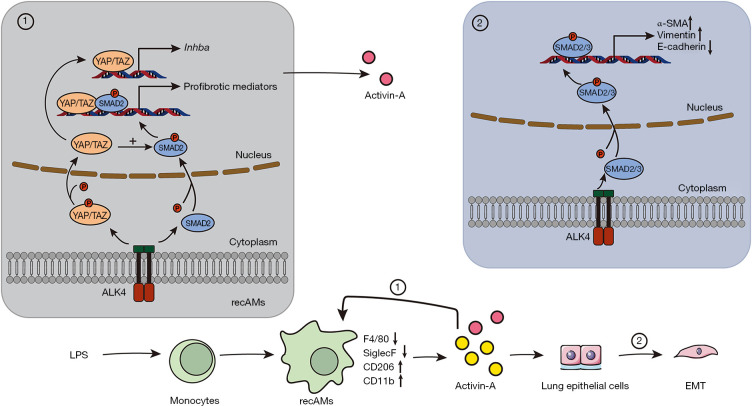
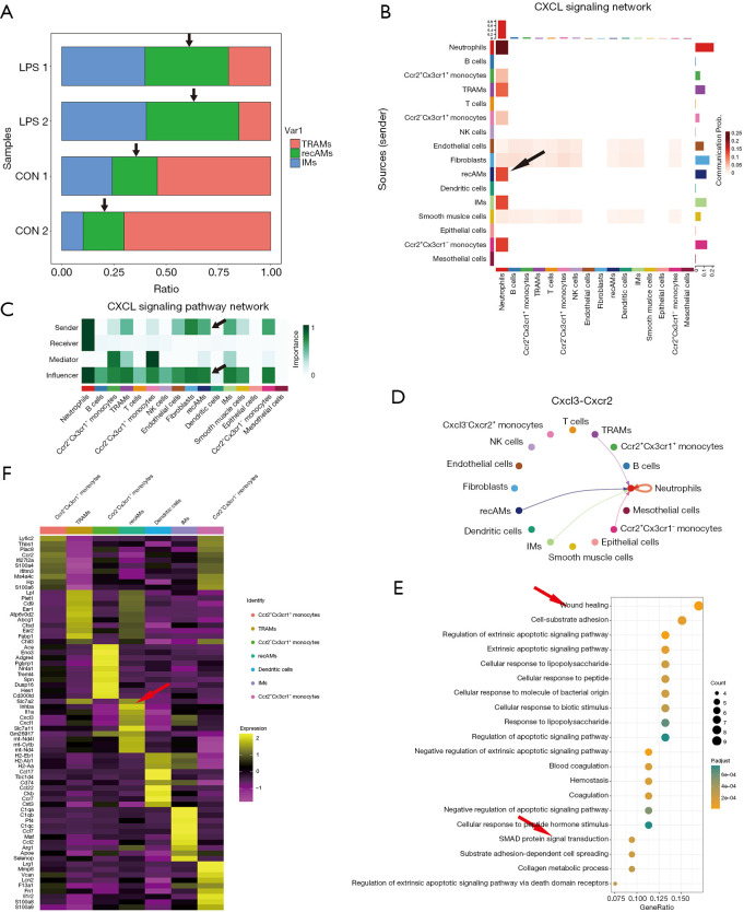
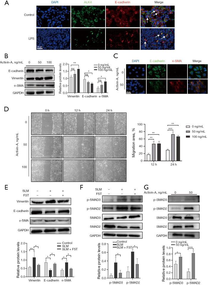
Our findings revealed that recAMs replaced tissue-resident alveolar macrophages (TRAMs) as the dominant macrophage population in the setting of ALI. The results of Gene Ontology (GO) analysis suggested that activin-A was associated with wound healing and suppressor of mothers against decapentaplegic (SMAD) protein signaling pathways. Immunofluorescence results revealed that the receptor of activin-A mainly localized to alveolar epithelial cells and macrophages. Subsequently, activin-A was specifically found to drive MLE-12 cells to mesenchymal cell transformation via the transforming growth factor-β (TGF-β)/SMAD signaling. Moreover, the results of transcriptome analysis and WB confirmed that activin-A could enhance the concerted activity of Hippo and TGF-β/SMAD pathways in BMDMs, leading to an increased expression of profibrotic mediator. Moreover, yes-associated protein (YAP) and transcriptional coactivated with PDZ-binding motif (TAZ) proteins were found to drive BMDM activin-A expression, which could generate a positive feedback mechanism that perpetuates fibrosis.

CONCLUSIONS

Our findings revealed that activin-A is involved in the pathological mechanisms in post-injury lung fibrosis by promoting epithelial-mesenchymal transition (EMT) and the formation of an underlying profibrotic positive feedback loop in recAMs. Activin-A is thus a potential therapeutic target for developing ALI and ALI-associated pulmonary fibrosis therapeutics.

摘要

背景

急性呼吸窘迫综合征(ARDS)是一种复杂的病理级联过程,其特征为肺部过度炎症反应和肺泡上皮细胞凋亡,最终导致呼吸功能障碍和衰竭。部分ARDS病例可发展为更为严重的肺纤维化状态,即损伤后肺纤维化。ARDS的死亡率和发病率较高,尤其是当它导致持续性肺泡和间质纤维化时,这需要紧急治疗和适当管理。脂多糖(LPS)诱导的急性肺损伤(ALI)小鼠模型已被广泛用于研究人类的ARDS。在我们的研究中,我们发现了这种小鼠模型中肺泡巨噬细胞(AM)谱的改变。具体而言,主要募集的AMs(recAMs)产生的激活素A被认为与损伤后肺纤维化过程有关。

方法

通过气管内注射3.5mg/kg LPS建立C57BL/6小鼠的ALI动物模型。单细胞RNA(scRNA)测序用于肺巨噬细胞的详细分类和功能表征。通过实验,我们使用酶联免疫吸附测定(ELISA)、组织学染色方法和免疫荧光评估激活素A在ALI小鼠模型损伤后肺纤维化中的作用。通过实验,我们使用蛋白质免疫印迹(WB)、定量实时聚合酶链反应、RNA测序和免疫荧光分析激活素A对小鼠肺上皮12(MLE-12)细胞和骨髓来源的巨噬细胞(BMDM)的影响。

结果

我们的研究结果表明,在ALI情况下,recAMs取代了组织驻留肺泡巨噬细胞(TRAMs)成为主要的巨噬细胞群体。基因本体论(GO)分析结果表明,激活素A与伤口愈合和母亲对脱磷酸化蛋白(SMAD)信号通路的抑制有关。免疫荧光结果显示,激活素A的受体主要定位于肺泡上皮细胞和巨噬细胞。随后,具体发现激活素A通过转化生长因子-β(TGF-β)/SMAD信号通路驱动MLE-12细胞向间充质细胞转化。此外,转录组分析和WB结果证实,激活素A可增强BMDM中Hippo和TGF-β/SMAD通路的协同活性,导致促纤维化介质表达增加。此外,发现Yes相关蛋白(YAP)和与PDZ结合基序(TAZ)共激活的转录蛋白可驱动BMDM激活素A的表达,并产生促进纤维化的正反馈机制。

结论

我们的研究结果表明,激活素A通过促进上皮-间质转化(EMT)和在recAMs中形成潜在的促纤维化正反馈环,参与损伤后肺纤维化的病理机制。因此,激活素A是开发ALI和ALI相关肺纤维化治疗方法的潜在治疗靶点。

https://cdn.ncbi.nlm.nih.gov/pmc/blobs/0212/11635239/422a33d5ec74/jtd-16-11-7709-f8.jpg
https://cdn.ncbi.nlm.nih.gov/pmc/blobs/0212/11635239/ca109c91d787/jtd-16-11-7709-f1.jpg
https://cdn.ncbi.nlm.nih.gov/pmc/blobs/0212/11635239/8d8b63c367a3/jtd-16-11-7709-f2.jpg
https://cdn.ncbi.nlm.nih.gov/pmc/blobs/0212/11635239/15c5169e96a0/jtd-16-11-7709-f3.jpg
https://cdn.ncbi.nlm.nih.gov/pmc/blobs/0212/11635239/c28ef006291c/jtd-16-11-7709-f4.jpg
https://cdn.ncbi.nlm.nih.gov/pmc/blobs/0212/11635239/dbfb26d0900d/jtd-16-11-7709-f5.jpg
https://cdn.ncbi.nlm.nih.gov/pmc/blobs/0212/11635239/5f661f422b89/jtd-16-11-7709-f6.jpg
https://cdn.ncbi.nlm.nih.gov/pmc/blobs/0212/11635239/49b431e17a76/jtd-16-11-7709-f7.jpg
https://cdn.ncbi.nlm.nih.gov/pmc/blobs/0212/11635239/422a33d5ec74/jtd-16-11-7709-f8.jpg

相似文献

1
Exacerbation of pulmonary fibrosis following acute lung injury via activin-A production by recruited alveolar macrophages.急性肺损伤后,募集的肺泡巨噬细胞通过产生激活素-A导致肺纤维化加重。
J Thorac Dis. 2024 Nov 30;16(11):7709-7728. doi: 10.21037/jtd-24-680. Epub 2024 Nov 14.
2
MBD2 promotes epithelial-to-mesenchymal transition (EMT) and ARDS-related pulmonary fibrosis by modulating FZD2.MBD2 通过调节 FZD2 促进上皮-间质转化(EMT)和急性呼吸窘迫综合征相关的肺纤维化。
Biochim Biophys Acta Mol Basis Dis. 2025 Jun;1871(5):167798. doi: 10.1016/j.bbadis.2025.167798. Epub 2025 Mar 12.
3
ISM1 suppresses LPS-induced acute lung injury and post-injury lung fibrosis in mice.ISM1 抑制 LPS 诱导的小鼠急性肺损伤和损伤后肺纤维化。
Mol Med. 2022 Jun 25;28(1):72. doi: 10.1186/s10020-022-00500-w.
4
Inhibition of EZH2 prevents acute respiratory distress syndrome (ARDS)-associated pulmonary fibrosis by regulating the macrophage polarization phenotype.抑制 EZH2 通过调节巨噬细胞极化表型预防急性呼吸窘迫综合征(ARDS)相关肺纤维化。
Respir Res. 2021 Jul 3;22(1):194. doi: 10.1186/s12931-021-01785-x.
5
[Effects of Hippo signaling pathway on lung injury repair by mesenchymal stem cells in acute respiratory distress syndrome].[河马信号通路对急性呼吸窘迫综合征中间充质干细胞肺损伤修复的影响]
Zhonghua Wei Zhong Bing Ji Jiu Yi Xue. 2019 Mar;31(3):281-287. doi: 10.3760/cma.j.issn.2095-4352.2019.03.005.
6
Depletion of circulating monocytes suppresses IL-17 and HMGB1 expression in mice with LPS-induced acute lung injury.循环单核细胞的耗竭可抑制脂多糖诱导的急性肺损伤小鼠中白细胞介素-17和高迁移率族蛋白B1的表达。
Am J Physiol Lung Cell Mol Physiol. 2017 Feb 1;312(2):L231-L242. doi: 10.1152/ajplung.00389.2016. Epub 2016 Dec 2.
7
APELIN-13 AMELIORATES LPS-INDUCED ENDOTHELIAL-TO-MESENCHYMAL TRANSITION AND POST-ACUTE LUNG INJURY PULMONARY FIBROSIS BY SUPPRESSING TRANSFORMING GROWTH FACTOR-Β1 SIGNALING.阿帕琳-13通过抑制转化生长因子-β1信号通路改善脂多糖诱导的内皮-间充质转化及急性肺损伤后肺纤维化
Shock. 2023 Jan 1;59(1):108-117. doi: 10.1097/SHK.0000000000002046. Epub 2022 Nov 15.
8
ATG16L1 restrains macrophage NLRP3 activation and alveolar epithelial cell injury during septic lung injury.自噬相关基因16样蛋白1(ATG16L1)在脓毒症肺损伤期间抑制巨噬细胞NLRP3激活及肺泡上皮细胞损伤。
Clin Transl Med. 2025 Apr;15(4):e70289. doi: 10.1002/ctm2.70289.
9
Keratinocyte growth factor expression is suppressed in early acute lung injury/acute respiratory distress syndrome by smad and c-Abl pathways.在早期急性肺损伤/急性呼吸窘迫综合征中,角质形成细胞生长因子的表达通过Smad和c-Abl信号通路受到抑制。
Crit Care Med. 2009 May;37(5):1678-84. doi: 10.1097/CCM.0b013e31819fc81a.
10
Lumican is elevated in the lung in human and experimental acute respiratory distress syndrome and promotes early fibrotic responses to lung injury.在人类和实验性急性呼吸窘迫综合征中,赖氨酰氧化酶在肺部升高,并促进肺损伤后早期的纤维化反应。
J Transl Med. 2022 Sep 4;20(1):392. doi: 10.1186/s12967-022-03597-z.

引用本文的文献

1
Activin A Inhibitory Peptides Suppress Fibrotic Pathways by Targeting Epithelial-Mesenchymal Transition and Fibroblast-Myofibroblast Transformation in Idiopathic Pulmonary Fibrosis.激活素A抑制肽通过靶向特发性肺纤维化中的上皮-间质转化和成纤维细胞-肌成纤维细胞转化来抑制纤维化途径。
Int J Mol Sci. 2025 Mar 17;26(6):2705. doi: 10.3390/ijms26062705.

本文引用的文献

1
Inhaled pirfenidone solution (AP01) for IPF: a randomised, open-label, dose-response trial.吸入性吡非尼酮溶液(AP01)治疗特发性肺纤维化:一项随机、开放标签、剂量反应试验。
Thorax. 2023 Sep;78(9):882-889. doi: 10.1136/thorax-2022-219391. Epub 2023 Mar 22.
2
MiR-302d inhibits TGFB-induced EMT and promotes MET in primary human RPE cells.miR-302d 抑制 TGFB 诱导的 EMT 并促进原代人 RPE 细胞中的 MET。
PLoS One. 2022 Nov 28;17(11):e0278158. doi: 10.1371/journal.pone.0278158. eCollection 2022.
3
Activin A and CCR2 regulate macrophage function in testicular fibrosis caused by experimental autoimmune orchitis.
激活素 A 和 CCR2 调节实验性自身免疫性睾丸炎引起的睾丸纤维化中的巨噬细胞功能。
Cell Mol Life Sci. 2022 Nov 24;79(12):602. doi: 10.1007/s00018-022-04632-4.
4
The Hippo pathway links adipocyte plasticity to adipose tissue fibrosis.Hippo 通路将脂肪细胞的可塑性与脂肪组织纤维化联系起来。
Nat Commun. 2022 Oct 13;13(1):6030. doi: 10.1038/s41467-022-33800-0.
5
Biology of lung macrophages in health and disease.肺巨噬细胞在健康和疾病中的生物学作用。
Immunity. 2022 Sep 13;55(9):1564-1580. doi: 10.1016/j.immuni.2022.08.010.
6
Pirfenidone and post-Covid-19 pulmonary fibrosis: invoked again for realistic goals.吡非尼酮与新冠后肺纤维化:再次为现实目标而呼吁。
Inflammopharmacology. 2022 Dec;30(6):2017-2026. doi: 10.1007/s10787-022-01027-6. Epub 2022 Aug 31.
7
Multiplatform Single-Cell Analysis Identifies Immune Cell Types Enhanced in Pulmonary Fibrosis.多平台单细胞分析鉴定出在肺纤维化中增强的免疫细胞类型。
Am J Respir Cell Mol Biol. 2022 Jul;67(1):50-60. doi: 10.1165/rcmb.2021-0418OC.
8
CD116+ fetal precursors migrate to the perinatal lung and give rise to human alveolar macrophages.CD116+ 胎儿前体细胞迁移到围生期肺,并产生人类肺泡巨噬细胞。
J Exp Med. 2022 Feb 7;219(2). doi: 10.1084/jem.20210987. Epub 2022 Jan 12.
9
The impact of the lung environment on macrophage development, activation and function: diversity in the face of adversity.肺部环境对巨噬细胞发育、激活和功能的影响:逆境中的多样性。
Mucosal Immunol. 2022 Feb;15(2):223-234. doi: 10.1038/s41385-021-00480-w. Epub 2022 Jan 11.
10
Progranulin and Activin A Concentrations are Elevated in Serum from Patients with Acute Exacerbations of Idiopathic Pulmonary Fibrosis.原肌球蛋白和激活素 A 浓度在特发性肺纤维化急性加重患者的血清中升高。
Lung. 2021 Oct;199(5):467-473. doi: 10.1007/s00408-021-00470-6. Epub 2021 Aug 30.