Ng Xiao Yu, Peh Gary, Morales-Wong Fernando, Gabriel Rami, Soong Poh Loong, Lin Kun-Han, Mehta Jodhbir S
Tissue Engineering and Cell Therapy Group, Singapore Eye Research Institute, Singapore 169856, Singapore.
Eye-Academic Clinical Program (ACP), Duke-National University of Singapore (NUS) Graduate Medical School, Singapore 169857, Singapore.
Cells. 2024 Dec 5;13(23):2012. doi: 10.3390/cells13232012.
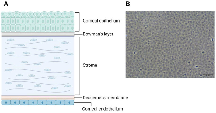
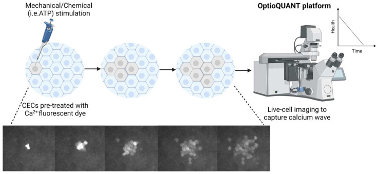
Corneal endothelium cells (CECs) regulate corneal hydration between the leaky barrier of the corneal endothelium and the ionic pumps on the surface of CECs. As CECs do not regenerate, loss of CECs leads to poor vision and corneal blindness. Corneal transplant is the only treatment option; however, there is a severe shortage of donor corneas globally. Cell therapy using propagated primary human CECs is an alternative approach to corneal transplantations, and proof of functionality is crucial for validating such CECs. Expression markers like Na-K-ATPase and ZO-1 are typical but not specific to CECs. Assessing the barrier function of the expanded CECs via electrical resistance (i.e., TEER and Ussing's chamber) involves difficult techniques and is thus impractical for clinical application. Calcium has been demonstrated to affect the paracellular permeability of the corneal endothelium. Its absence alters morphology and disrupts apical junctions in bovine CECs, underscoring its importance. Calcium signaling patterns such as calcium waves affect the rate of wound healing in bovine CECs. Therefore, observing calcium waves in expanded CECs could provide valuable insights into their health and functional integrity. Mechanical or chemical stimulations, combined with Ca-sensitive fluorescent dyes and time-lapse imaging, can be used to visualize these waves, which could potentially be used to qualify expanded CECs.
角膜内皮细胞(CECs)在角膜内皮的渗漏屏障和CECs表面的离子泵之间调节角膜水合作用。由于CECs不会再生,CECs的丧失会导致视力下降和角膜失明。角膜移植是唯一的治疗选择;然而,全球范围内供体角膜严重短缺。使用扩增的原代人CECs进行细胞治疗是角膜移植的一种替代方法,而功能验证对于确认此类CECs至关重要。诸如钠钾ATP酶和紧密连接蛋白1(ZO-1)等表达标志物是典型的,但并非CECs所特有。通过电阻(即跨上皮电阻和尤斯灌流小室)评估扩增的CECs的屏障功能涉及复杂的技术,因此在临床应用中不切实际。钙已被证明会影响角膜内皮的细胞旁通透性。其缺失会改变牛CECs的形态并破坏顶端连接,凸显了其重要性。诸如钙波等钙信号模式会影响牛CECs的伤口愈合速度。因此,观察扩增的CECs中的钙波可以为其健康状况和功能完整性提供有价值的见解。机械或化学刺激,结合钙敏荧光染料和延时成像,可用于可视化这些波,这有可能用于鉴定扩增的CECs。