Chaphalkar Renuka M, Kodati Bindu, Maddineni Prabhavathi, He Shaoqing, Brooks Calvin D, Stankowska Dorota L, Yang Shaohua, Zode Gulab, Krishnamoorthy Raghu R
Department of Pharmacology and Neuroscience, University of North Texas Health Science Center, Fort Worth, TX 76107, USA.
North Texas Eye Research Institute, University of North Texas Health Science Center, Fort Worth, TX 76107, USA.
Int J Mol Sci. 2024 Dec 4;25(23):13040. doi: 10.3390/ijms252313040.
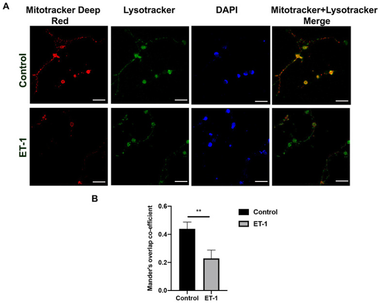
Glaucoma is a heterogenous group of optic neuropathies characterized by the degeneration of optic nerve axons and the progressive loss of retinal ganglion cells (RGCs), which could ultimately lead to vision loss. Elevated intraocular pressure (IOP) is a major risk factor in the development of glaucoma, and reducing IOP remains the main therapeutic strategy. Endothelin-1 (ET-1), a potent vasoactive peptide, has been shown to produce neurodegenerative effects in animal models of glaucoma. However, the detailed mechanisms underlying ET-1-mediated neurodegeneration in glaucoma are not completely understood. In the current study, using a Seahorse Mitostress assay, we report that ET-1 treatment for 4 h and 24 h time points causes a significant decline in various parameters of mitochondrial function, including ATP production, maximal respiration, and spare respiratory capacity in cultured RGCs. This compromise in mitochondrial function could trigger activation of mitophagy as a quality control mechanism to restore RGC health. Contrary to our expectation, we observed a decrease in mitophagy following ET-1 treatment for 24 h in cultured RGCs. Using Morrison's model of ocular hypertension in rats, we investigated here, for the first time, changes in mitophagosome formation by analyzing the co-localization of LC-3B and TOM20 in RGCs. We also injected ET-1 (24 h) into transgenic GFP-LC3 mice to analyze the formation of mitophagosomes in vivo. In Morrison's model of ocular hypertension, as well as in ET-1 injected GFP-LC3 mice, we found a decrease in co-localization of LC3 and TOM20, indicating reduced mitophagy. Taken together, these results demonstrate that both ocular hypertension and ET-1 administration in rats and mice lead to reduced mitophagy, thus predisposing RGCs to neurodegeneration.
青光眼是一组异质性视神经病变,其特征是视神经轴突退化和视网膜神经节细胞(RGCs)逐渐丧失,最终可能导致视力丧失。眼压升高是青光眼发展的主要危险因素,降低眼压仍然是主要的治疗策略。内皮素-1(ET-1)是一种强效血管活性肽,已被证明在青光眼动物模型中产生神经退行性作用。然而,ET-1介导的青光眼神经退行性变的详细机制尚未完全了解。在本研究中,我们使用海马体线粒体应激试验报告,ET-1处理4小时和24小时时间点会导致培养的RGCs线粒体功能的各种参数显著下降,包括ATP产生、最大呼吸和备用呼吸能力。线粒体功能的这种损害可能触发线粒体自噬作为一种质量控制机制来恢复RGC的健康。与我们的预期相反,我们观察到在培养的RGCs中ET-1处理24小时后线粒体自噬减少。我们使用大鼠高眼压的莫里森模型,首次通过分析RGCs中LC-3B和TOM20的共定位来研究线粒体自噬体形成的变化。我们还将ET-1(24小时)注射到转基因GFP-LC3小鼠中,以分析体内线粒体自噬体的形成。在莫里森高眼压模型以及注射ET-1的GFP-LC3小鼠中,我们发现LC3和TOM20的共定位减少,表明线粒体自噬减少。综上所述,这些结果表明,大鼠和小鼠的高眼压和ET-1给药均导致线粒体自噬减少,从而使RGCs易发生神经退行性变。