Kooner Kiren, Rubiños Carlos
Southfields Veterinary Specialists (Part of Linnaeus Veterinary Limited), Basildon, UK.
JFMS Open Rep. 2024 Dec 16;10(2):20551169241285257. doi: 10.1177/20551169241285257. eCollection 2024 Jul-Dec.
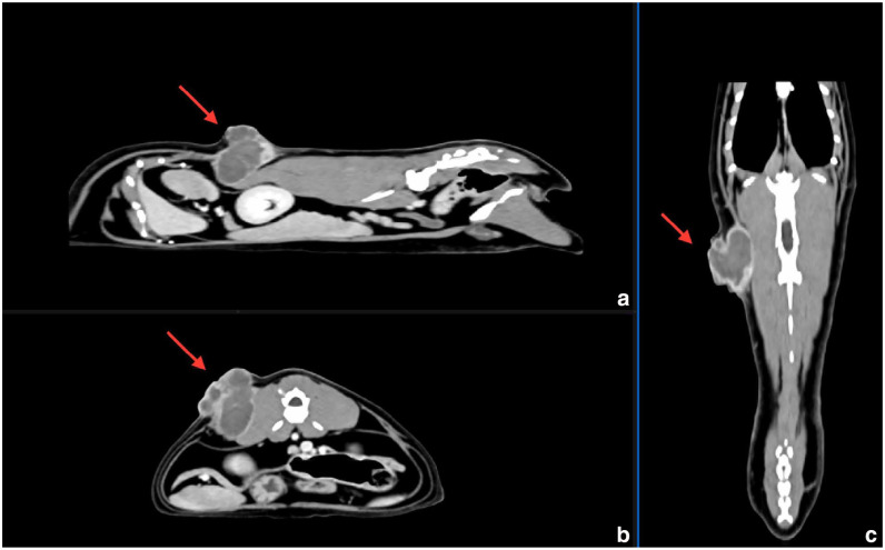
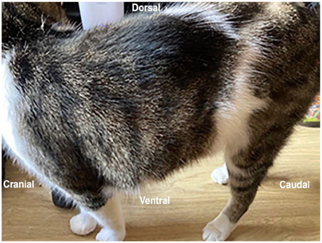
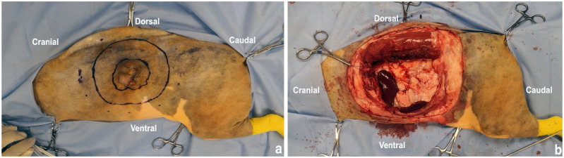
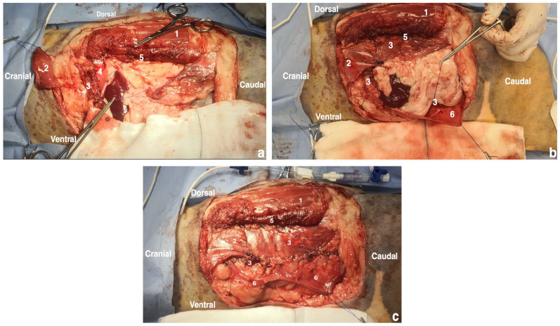
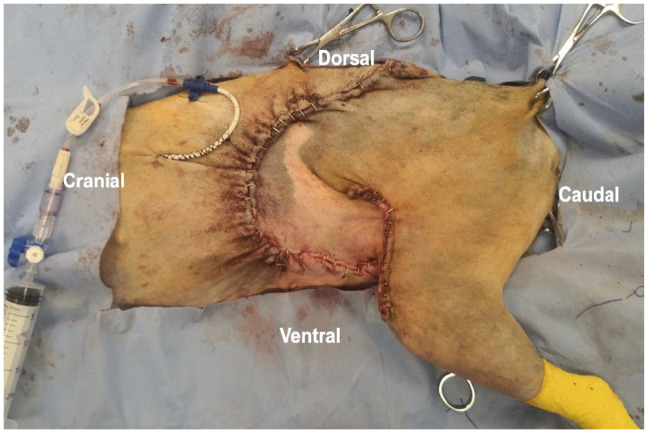
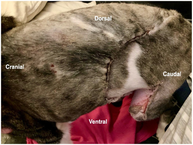
A cat aged 12 years and 7 months was referred to a multidisciplinary hospital for investigation of feline injection site sarcoma (FISS) on the left thoracolumbar region. A CT examination of the mass revealed a multi-lobulated mass affecting the body wall, extending from the level of lumbar vertebrae L2 to L4. The mass was excised with 5 cm lateral margins, including resection of the 13th left rib, the caudal edge of the latissimus dorsi (LD) muscle, full-thickness abdominal wall and sections of the lumbar epaxial muscles. To reconstruct the defect, a combination of muscle flaps was used. This included diaphragmatic advancement and lateralisation, rotation of the LD, and creation of transposition flaps from the internal abdominal oblique and external abdominal oblique muscles, ensuring closure without tension. Skin closure required mobilising an inguinal flank fold flap. The cat was discharged from hospital 3 days postoperatively. Histopathology confirmed a diagnosis of FISS with clean wide margins. A gradual return to normal activity and complete healing of the surgical site was reported on follow-up, with one minor complication related to the skin flap (bruising at the base of the inguinal flank fold flap).
This report describes the use of the aforementioned combination of muscle flaps to close a major abdominal wall defect in a cat with an excellent outcome. Practitioners can consider this technique when planning tissue reconstruction after FISS resection.
一只12岁7个月大的猫被转诊至一家多学科医院,以检查左胸腰段的猫注射部位肉瘤(FISS)。对肿块进行的CT检查显示,一个多叶状肿块累及体壁,从腰椎L2水平延伸至L4。肿块以5厘米的外侧切缘切除,包括切除第13根左肋骨、背阔肌(LD)的后缘、全层腹壁和部分腰轴肌。为了修复缺损,使用了多种肌皮瓣组合。这包括膈肌推进和外移、LD旋转,以及利用腹内斜肌和腹外斜肌制作转位皮瓣,确保无张力缝合。皮肤缝合需要游离一个腹股沟侧翼折叠皮瓣。这只猫在术后3天出院。组织病理学确诊为FISS,切缘干净且广泛。随访报告显示,猫逐渐恢复正常活动,手术部位完全愈合,仅出现了一个与皮瓣相关的轻微并发症(腹股沟侧翼折叠皮瓣底部出现瘀伤)。
本报告描述了使用上述肌皮瓣组合修复一只猫的主要腹壁缺损,取得了良好效果。从业者在计划FISS切除术后的组织重建时可考虑该技术。