Chen Jian, Li Wei, Zhang Cheng, Wen Dihao, Jiao Cheng
Department of General Surgery, Bethune International Peace Hospital of The People's Liberation Army, No. 398, Zhongshan XI Road, Qiaoxi District, Shijiazhuang, 050000, Hebei, People's Republic of China.
Department of Gastroenterology, Bethune International Peace Hospital of The People's Liberation Army, Shijiazhuang, 050000, Hebei, People's Republic of China.
Discov Oncol. 2024 Dec 18;15(1):793. doi: 10.1007/s12672-024-01586-w.
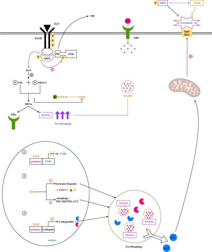
To elucidate the mechanism by which tyrosine phosphatase SHP2 protects CRC through modulation of TFEB-mediated ferritinophagy, thereby suppressing ROS and ferroptosis.
SW480 and SW620 cells, in the logarithmic growth phase, were treated with or without the SHP2 inhibitor PHPS1, the activator Trichomide A, EGF, or MMP inhibitors, and randomly assigned to four groups. Additionally, SW480 cells in the logarithmic phase underwent treatments with EGF, the ferroptosis inducer erastin, Trichomide A, or the curcumin analog C1, forming seven groups. Cell migration assessment in these groups employed scratch and Transwell assays. Protein expression analysis of total SHP2, total PI3K, p-SHP2, p-PI3K, p-TFEB, TFEB, SQSTM1, LC3, LAMP2, NCOA4, FTH1, GPX4, NOX4, and ACSL4 in the seven SW480 groups was conducted through Western blot and immunofluorescence. Apoptosis analysis was performed on these seven groups, while gene co-expression analysis utilized bioinformatics. SW480 and CCD-841CoN cells were categorized into four groups, undergoing treatment with saline, EGFR-OE lentivirus, SHP2-KD lentivirus, or SHP2-OE lentivirus. Western blot analysis in SW480 cells detected EGFR, total SHP2, p-SHP2, GPX4, and ACSL4 proteins, and tumor volume observations were conducted in a nude mouse xenograft model. Western blot also evaluated total SHP2, p-SHP2, GPX4, and ACSL4 protein expression in CCD-841CoN cells.
Bioinformatics analysis revealed correlations between EGFR and SHP2, SHP2 and PIK3CA, SHP2 and MAPK1, BRK4 and HIF1A, HIF1A and NCOA4, as well as TFEB and FTH1. Scratch and Transwell assays showed that SHP2 diminishes the migratory capacity of SW480 and SW620 cells. Western blot and immunofluorescence demonstrated that EGFR activation of SHP2 markedly elevated p-TFEB levels while reducing TFEB protein expression. EGF stimulation enhanced the expression of FTH1, GPX4, NOX4, and ACSL4. Combined stimulation with EGF and SHP2 further amplified the expression of p-SHP2, p-TFEB, and NCOA4 while reducing TFEB, SQSTM1, LC3, and LAMP2. Erastin augmented FTH1, GPX4, NOX4, and ACSL4 expression while decreasing p-SHP2, p-TFEB, TFEB, SQSTM1, LC3, LAMP2, and NCOA4. TFEB activation suppressed p-SHP2, p-TFEB, NCOA4, FTH1, and GPX4 expression, while promoting TFEB, SQSTM1, LC3, LAMP2, NOX4, and ACSL4 expression. Apoptosis assays indicated that SHP2 activation decelerated apoptosis in SW480 cells, whereas erastin under EGF stimulation accelerated apoptosis, as did TFEB activation. Western blot results in SW480 cells displayed that overexpression of EGFR or SHP2 significantly increased total SHP2, p-SHP2, and GPX4 expression while decreasing ACSL4 levels. SHP2 knockdown decreased total SHP2, p-SHP2, and GPX4 expression, with an increase in ACSL4 expression. In CCD-841CoN cells, overexpression of EGFR or SHP2 resulted in a decrease in p-SHP2 and an increase in total SHP2, more pronounced with SHP2 overexpression, while GPX4 and ACSL4 levels remained stable. SHP2 knockdown led to reduced EGFR, total SHP2, p-SHP2, and GPX4 expression, without a significant impact on ACSL4 levels. The nude mouse xenograft model demonstrated that EGFR overexpression significantly increased tumor size, whereas SHP2 overexpression markedly decreased tumor volume. SHP2 knockdown resulted in significantly larger tumors.
SHP2 advances CRC progression by modulating TFEB-mediated ferritinophagy, suppressing ROS and ferroptosis. Targeting SHP2 presents a promising therapeutic strategy for CRC.
阐明酪氨酸磷酸酶SHP2通过调节TFEB介导的铁蛋白自噬来保护结直肠癌的机制,从而抑制活性氧(ROS)和铁死亡。
处于对数生长期的SW480和SW620细胞,分别用或不用SHP2抑制剂PHPS1、激活剂曲古抑菌素A、表皮生长因子(EGF)或基质金属蛋白酶(MMP)抑制剂处理,并随机分为四组。此外,对数期的SW480细胞用EGF、铁死亡诱导剂厄洛替尼、曲古抑菌素A或姜黄素类似物C1处理,形成七组。这些组中的细胞迁移评估采用划痕和Transwell实验。通过蛋白质免疫印迹法和免疫荧光法对七个SW480组中的总SHP2、总磷脂酰肌醇-3激酶(PI3K)、磷酸化SHP2(p-SHP2)、磷酸化PI3K(p-PI3K)、磷酸化转录因子EB(p-TFEB)、TFEB、p62、微管相关蛋白1轻链3(LC3)、溶酶体相关膜蛋白2(LAMP2)、核受体辅助激活因子4(NCOA4)、铁蛋白重链1(FTH1)、谷胱甘肽过氧化物酶4(GPX4)、烟酰胺腺嘌呤二核苷酸磷酸氧化酶4(NOX4)和长链脂酰辅酶A合成酶4(ACSL4)进行蛋白质表达分析。对这七组进行凋亡分析,同时利用生物信息学进行基因共表达分析。SW480和CCD-841CoN细胞分为四组,分别用生理盐水、表皮生长因子受体(EGFR)过表达慢病毒、SHP2基因敲低慢病毒或SHP2过表达慢病毒处理。对SW480细胞进行蛋白质免疫印迹分析,检测EGFR、总SHP2、p-SHP2、GPX4和ACSL4蛋白,并在裸鼠异种移植模型中观察肿瘤体积。蛋白质免疫印迹法还评估了CCD-841CoN细胞中总SHP2、p-SHP2、GPX4和ACSL4蛋白的表达。
生物信息学分析显示EGFR与SHP2、SHP2与PIK3CA、SHP2与丝裂原活化蛋白激酶1(MAPK1)、BRK4与缺氧诱导因子1α(HIF1A)、HIF1A与NCOA4以及TFEB与FTH1之间存在相关性。划痕和Transwell实验表明,SHP2降低了SW480和SW620细胞的迁移能力。蛋白质免疫印迹法和免疫荧光法表明,EGFR激活SHP2显著提高了p-TFEB水平,同时降低了TFEB蛋白表达。EGF刺激增强了FTH1、GPX4、NOX4和ACSL4的表达。EGF和SHP2联合刺激进一步放大了p-SHP2、p-TFEB和NCOA4的表达,同时降低了TFEB、p62、LC3和LAMP2的表达。厄洛替尼增加了FTH1、GPX4、NOX4和ACSL4的表达,同时降低了p-SHP2、p-TFEB、TFEB、p62、LC3、LAMP2和NCOA4的表达。TFEB激活抑制了p-SHP2、p-TFEB、NCOA4、FTH1和GPX4的表达,同时促进了TFEB、p62、LC3、LAMP2、NOX4和ACSL4的表达。凋亡分析表明,SHP2激活减缓了SW480细胞的凋亡,而在EGF刺激下厄洛替尼加速了凋亡,TFEB激活也有同样的效果。SW480细胞的蛋白质免疫印迹结果显示,EGFR或SHP2过表达显著增加了总SHP2、p-SHP2和GPX4的表达,同时降低了ACSL4水平。SHP2基因敲低降低了总SHP2、p-SHP2和GPX4的表达,同时ACSL4表达增加。在CCD-841CoN细胞中,EGFR或SHP2过表达导致p-SHP2降低,总SHP2增加,SHP2过表达时更明显,而GPX4和ACSL4水平保持稳定。SHP2基因敲低导致EGFR、总SHP2、p-SHP2和GPX4表达降低,对ACSL4水平无显著影响。裸鼠异种移植模型表明,EGFR过表达显著增加了肿瘤大小,而SHP2过表达显著降低了肿瘤体积。SHP2基因敲低导致肿瘤显著增大。
SHP2通过调节TFEB介导的铁蛋白自噬、抑制ROS和铁死亡促进结直肠癌进展。靶向SHP2为结直肠癌提供了一种有前景的治疗策略。