• 文献检索
  • 文档翻译
  • 深度研究
  • 学术资讯
  • Suppr Zotero 插件Zotero 插件
  • 邀请有礼
  • 套餐&价格
  • 历史记录
应用&插件
Suppr Zotero 插件Zotero 插件浏览器插件Mac 客户端Windows 客户端微信小程序
定价
高级版会员购买积分包购买API积分包
服务
文献检索文档翻译深度研究API 文档MCP 服务
关于我们
关于 Suppr公司介绍联系我们用户协议隐私条款
关注我们

Suppr 超能文献

核心技术专利:CN118964589B侵权必究
粤ICP备2023148730 号-1Suppr @ 2026

文献检索

告别复杂PubMed语法,用中文像聊天一样搜索,搜遍4000万医学文献。AI智能推荐,让科研检索更轻松。

立即免费搜索

文件翻译

保留排版,准确专业,支持PDF/Word/PPT等文件格式,支持 12+语言互译。

免费翻译文档

深度研究

AI帮你快速写综述,25分钟生成高质量综述,智能提取关键信息,辅助科研写作。

立即免费体验

MRI在检测甲状腺乳头状癌甲状腺外侵犯中不断演变的作用:一项系统评价和荟萃分析

The evolving role of MRI in the detection of extrathyroidal extension of papillary thyroid carcinoma: a systematic review and meta-analysis.

作者信息

Javid Mona, Mirdamadi Arian, Javid Mohammadreza, Keivanlou Mohammad-Hossein, Amini-Salehi Ehsan, Norouzi Naeim, Abbaspour Elahe, Alizadeh Ahmad, Joukar Farahnaz, Mansour-Ghanaei Fariborz, Hassanipour Soheil

机构信息

Gastrointestinal and Liver Diseases Research Center, Guilan University of Medical Sciences, Rasht, Iran.

Student Research Committee, School of Medicine, Guilan University of Medical Sciences, Rasht, Iran.

出版信息

BMC Cancer. 2024 Dec 18;24(1):1531. doi: 10.1186/s12885-024-13288-1.

DOI:10.1186/s12885-024-13288-1
PMID:39696088
原文链接:https://pmc.ncbi.nlm.nih.gov/articles/PMC11657418/
Abstract

BACKGROUND

Papillary thyroid carcinoma (PTC) is the predominant form of thyroid cancer, and the presence of extrathyroidal extension (ETE) significantly impacts treatment decisions and prognosis. Accurate preoperative detection of ETE remains challenging, highlighting the need to evaluate advanced imaging techniques.This systematic review and meta-analysis aimed to investigate the diagnostic accuracy of magnetic resonance imaging (MRI) in detecting extrathyroidal extension (ETE) among patients diagnosed with papillary thyroid carcinoma (PTC).

METHOD

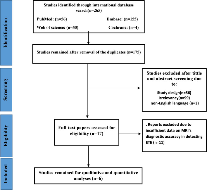
We conducted a comprehensive search of global databases including PubMed, Web of Science, EMBASE, and the Cochrane Library, spanning from inception to November 03, 2024. We included studies that utilized preoperative MRI to evaluate the presence of ETE. Quality assessment was carried out using the Joanna Briggs Institute (JBI) standard checklists. Data analysis was performed using Comprehensive Meta-Analysis (CMA) software version 3. The study protocol was registered in PROSPERO (CRD42024499536).

RESULT

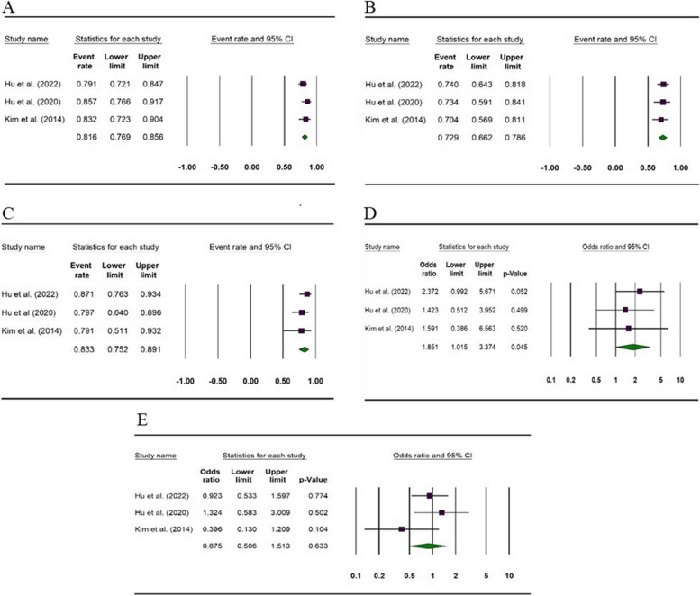
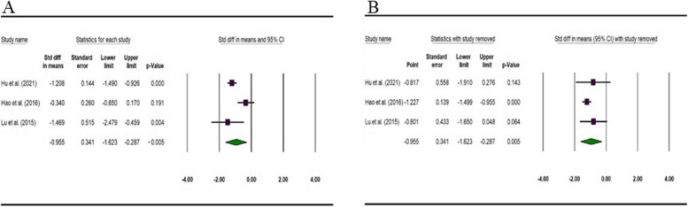
Six studies were included in our final quantitative analysis. The included studies were classified into two groups; the first group focused on evaluating the accuracy of MRI in detecting ETE, while the second group assessed the apparent diffusion coefficient (ADC). The accuracy of MRI for overall ETE, minimal ETE (mETE), and gross ETE (gETE) was 81.0% (95% CI: 76.9%-85.6%), 72.9% (95% CI: 66.2%-78.6%), and 83.3% (95% CI: 75.2%-89.1%), respectively. MRI demonstrated a statistically significant difference in detecting gETE compared to mETE (OR = 1.85, 95% CI: 1.01-3.37, P-value = 0.045). Our analysis showed that the ADC of the lesion for b-value 500 is lower in patients with ETE compared to those without ETE (SMD = 0.95, 95% CI: 0.28-1.62, P-value = 0.005).

CONCLUSION

Our findings demonstrate that MRI has substantial accuracy in detecting ETE in PTC, especially for gross ETE. This suggests MRI could be a valuable tool in preoperative planning, helping to guide surgical decision-making by more precisely identifying patients at higher risk. However, the limited number of studies underscores the need for further research to confirm MRI's role in routine clinical practice and to refine imaging protocols for more accurate differentiation between minimal and gross ETE.

摘要

背景

乳头状甲状腺癌(PTC)是甲状腺癌的主要形式,甲状腺外侵犯(ETE)的存在显著影响治疗决策和预后。术前准确检测ETE仍然具有挑战性,这凸显了评估先进成像技术的必要性。本系统评价和荟萃分析旨在研究磁共振成像(MRI)在诊断为乳头状甲状腺癌(PTC)的患者中检测甲状腺外侵犯(ETE)的诊断准确性。

方法

我们对包括PubMed、Web of Science、EMBASE和Cochrane图书馆在内的全球数据库进行了全面检索,检索时间跨度从建库至2024年11月3日。我们纳入了利用术前MRI评估ETE存在情况的研究。使用乔安娜·布里格斯研究所(JBI)标准清单进行质量评估。使用综合荟萃分析(CMA)软件版本3进行数据分析。该研究方案已在PROSPERO(CRD42024499536)中注册。

结果

六项研究纳入了我们最终的定量分析。纳入的研究分为两组;第一组侧重于评估MRI检测ETE的准确性,而第二组评估表观扩散系数(ADC)。MRI对总体ETE、微小ETE(mETE)和大体ETE(gETE)的检测准确性分别为81.0%(95%CI:76.9%-85.6%)、72.9%(95%CI:66.2%-78.6%)和83.3%(95%CI:75.2%-89.1%)。与mETE相比,MRI在检测gETE方面显示出统计学显著差异(OR = 1.85,95%CI:1.01-3.37,P值 = 0.045)。我们的分析表明,与无ETE的患者相比,有ETE的患者病变在b值为500时的ADC较低(标准化均数差 = 0.95,95%CI:0.28-1.62,P值 = 0.005)。

结论

我们的研究结果表明,MRI在检测PTC中的ETE方面具有较高的准确性,尤其是对于大体ETE。这表明MRI可能是术前规划中的一个有价值的工具,通过更精确地识别高风险患者来帮助指导手术决策。然而,研究数量有限强调了需要进一步研究以确认MRI在常规临床实践中的作用,并完善成像方案以更准确地区分微小和大体ETE。

https://cdn.ncbi.nlm.nih.gov/pmc/blobs/895b/11657418/0b0f25b16890/12885_2024_13288_Fig5_HTML.jpg
https://cdn.ncbi.nlm.nih.gov/pmc/blobs/895b/11657418/8e1872b3554e/12885_2024_13288_Fig1_HTML.jpg
https://cdn.ncbi.nlm.nih.gov/pmc/blobs/895b/11657418/a0f1d316c05f/12885_2024_13288_Fig2_HTML.jpg
https://cdn.ncbi.nlm.nih.gov/pmc/blobs/895b/11657418/302a7859b3c9/12885_2024_13288_Fig3_HTML.jpg
https://cdn.ncbi.nlm.nih.gov/pmc/blobs/895b/11657418/be86549f4ae1/12885_2024_13288_Fig4_HTML.jpg
https://cdn.ncbi.nlm.nih.gov/pmc/blobs/895b/11657418/0b0f25b16890/12885_2024_13288_Fig5_HTML.jpg

相似文献

1
The evolving role of MRI in the detection of extrathyroidal extension of papillary thyroid carcinoma: a systematic review and meta-analysis.MRI在检测甲状腺乳头状癌甲状腺外侵犯中不断演变的作用:一项系统评价和荟萃分析
BMC Cancer. 2024 Dec 18;24(1):1531. doi: 10.1186/s12885-024-13288-1.
2
Is There a Difference Between Minimal and Gross Extension into the Strap Muscles for the Risk of Recurrence in Papillary Thyroid Carcinomas?微小和广泛侵犯胸锁乳突肌对甲状腺乳头状癌复发风险的影响是否存在差异?
Thyroid. 2020 Jul;30(7):1008-1016. doi: 10.1089/thy.2019.0753. Epub 2020 Mar 17.
3
Preoperative assessment of extrathyroidal extension of papillary thyroid carcinomas by ultrasound and magnetic resonance imaging: a comparative study.超声与磁共振成像对甲状腺外侵犯的甲状腺乳头状癌术前评估的对比研究。
Radiol Med. 2020 Sep;125(9):870-876. doi: 10.1007/s11547-020-01184-0. Epub 2020 Apr 5.
4
Preoperative prediction of the extrathyroidal extension of papillary thyroid carcinoma with ultrasonography versus MRI: a retrospective cohort study.术前超声与 MRI 对甲状腺癌甲状腺外侵犯的预测:一项回顾性队列研究。
Int J Surg. 2014;12(5):544-8. doi: 10.1016/j.ijsu.2014.03.003. Epub 2014 Mar 12.
5
The Diagnostic Performance of Ultrasonography in the Evaluation of Extrathyroidal Extension in Papillary Thyroid Carcinoma: A Systematic Review and Meta-Analysis.超声检查在评估甲状腺乳头状癌甲状腺外侵犯中的诊断性能:系统评价和荟萃分析。
Int J Mol Sci. 2022 Dec 26;24(1):371. doi: 10.3390/ijms24010371.
6
[Diagnostic value of combination of US and MRI in preoperative prediction for the extrathyroidal extension of papillary thyroid carcinoma].[超声与磁共振成像联合应用在术前预测甲状腺乳头状癌甲状腺外侵犯中的诊断价值]
Zhonghua Yi Xue Za Zhi. 2017 Sep 5;97(33):2600-2603. doi: 10.3760/cma.j.issn.0376-2491.2017.33.010.
7
Accuracy of Radiomics in the Identification of Extrathyroidal Extension and BRAF Mutations in Papillary Thyroid Carcinoma: A Systematic Review and Meta-analysis.放射组学在识别甲状腺乳头状癌甲状腺外侵犯和BRAF突变中的准确性:一项系统评价和荟萃分析
Acad Radiol. 2025 Mar;32(3):1385-1397. doi: 10.1016/j.acra.2024.11.014. Epub 2025 Jan 6.
8
Can texture analysis of T2-weighted MRI be used to predict extrathyroidal extension in papillary thyroid carcinoma?T2 加权 MRI 的纹理分析能否用于预测甲状腺癌的甲状腺外侵犯?
Medicine (Baltimore). 2023 Nov 3;102(44):e35800. doi: 10.1097/MD.0000000000035800.
9
Prognostic impact of minimal extrathyroidal extension in papillary thyroid carcinoma.甲状腺乳头状癌中最小甲状腺外侵犯的预后影响
Medicine (Baltimore). 2016 Dec;95(52):e5794. doi: 10.1097/MD.0000000000005794.
10
Using diffusion-weighted MRI to predict aggressive histological features in papillary thyroid carcinoma: a novel tool for pre-operative risk stratification in thyroid cancer.使用扩散加权磁共振成像预测甲状腺乳头状癌的侵袭性组织学特征:一种用于甲状腺癌术前风险分层的新工具。
Thyroid. 2015 Jun;25(6):672-80. doi: 10.1089/thy.2014.0419. Epub 2015 Apr 21.

本文引用的文献

1
Macrofollicular subtype of papillary thyroid carcinoma: Ultrasonographic findings and clinical implications.甲状腺乳头状癌的大滤泡亚型:超声表现及临床意义。
Medicine (Baltimore). 2024 Oct 4;103(40):e39944. doi: 10.1097/MD.0000000000039944.
2
Role of CT imaging in addressing resectability issues in differentiated thyroid cancer: imaging-based Mahajan grading system for TI and ETE.CT成像在解决分化型甲状腺癌可切除性问题中的作用:基于成像的TI和ETE的马氏分级系统
Front Oncol. 2024 Sep 13;14:1382518. doi: 10.3389/fonc.2024.1382518. eCollection 2024.
3
The superior value of radiomics to sonographic assessment for ultrasound-based evaluation of extrathyroidal extension in papillary thyroid carcinoma: a retrospective study.
基于超声的甲状腺外侵犯评估中,放射组学优于超声评估:一项回顾性研究。
Radiol Oncol. 2024 Sep 15;58(3):386-396. doi: 10.2478/raon-2024-0040. eCollection 2024 Sep 1.
4
The value of apparent diffusion coefficient values in predicting Gleason grading of low to intermediate-risk prostate cancer.表观扩散系数值在预测低至中危前列腺癌Gleason分级中的价值。
Insights Imaging. 2024 Jun 9;15(1):137. doi: 10.1186/s13244-024-01684-x.
5
Can texture analysis of T2-weighted MRI be used to predict extrathyroidal extension in papillary thyroid carcinoma?T2 加权 MRI 的纹理分析能否用于预测甲状腺癌的甲状腺外侵犯?
Medicine (Baltimore). 2023 Nov 3;102(44):e35800. doi: 10.1097/MD.0000000000035800.
6
Factors influencing extrathyroidal extension of papillary thyroid cancer and evaluation of ultrasonography for its diagnosis: a retrospective analysis.影响甲状腺外侵犯的因素及超声对此的诊断价值:回顾性分析。
Sci Rep. 2023 Oct 26;13(1):18344. doi: 10.1038/s41598-023-45642-x.
7
The Diagnostic Performance of Ultrasonography in the Evaluation of Extrathyroidal Extension in Papillary Thyroid Carcinoma: A Systematic Review and Meta-Analysis.超声检查在评估甲状腺乳头状癌甲状腺外侵犯中的诊断性能:系统评价和荟萃分析。
Int J Mol Sci. 2022 Dec 26;24(1):371. doi: 10.3390/ijms24010371.
8
The Role of Diffusion-Weighted Magnetic Resonance Imaging in Differentiating Benign From Malignant Thyroid Nodules: A Descriptive Observational Study.扩散加权磁共振成像在鉴别甲状腺良恶性结节中的作用:一项描述性观察研究。
Cureus. 2022 Oct 19;14(10):e30493. doi: 10.7759/cureus.30493. eCollection 2022 Oct.
9
"Micro" Extrathyroidal Extension in Risk Stratification for Papillary Thyroid Carcinoma: Should It Be in the Intermediate-Risk or High-Risk Group? A Single-Center Retrospective Study.甲状腺乳头状癌风险分层中的“微小”甲状腺外侵犯:应归为中危组还是高危组?一项单中心回顾性研究
Cancer Manag Res. 2022 Nov 16;14:3181-3190. doi: 10.2147/CMAR.S390468. eCollection 2022.
10
Thyroid Carcinoma, Version 2.2022, NCCN Clinical Practice Guidelines in Oncology.甲状腺癌临床实践指南(NCCN 指南)2022 年第 2 版。
J Natl Compr Canc Netw. 2022 Aug;20(8):925-951. doi: 10.6004/jnccn.2022.0040.