Wu Chunxue, Dong Mengqi, Zang Zhenxiang, Shan Yi, Yu Jiaxing, Hong Tao, Yang Kun, Zhao Cheng, Zhang Hongqi, Lu Jie
Department of Radiology and Nuclear Medicine, Xuanwu Hospital, Capital Medical University, Beijing, China.
Beijing Key Laboratory of Magnetic Resonance Imaging and Brain Informatics, Beijing, China.
Quant Imaging Med Surg. 2024 Dec 5;14(12):8974-8987. doi: 10.21037/qims-24-1097. Epub 2024 Nov 29.
Preliminary small-sample studies suggest that silent magnetic resonance angiography (MRA) has an advantage over time-of-flight MRA (TOF MRA) in the characterization of brain arteriovenous malformation (BAVM), but did not examine whether the imaging performance of silent MRA was affected by the intrinsic features of BAVM or common clinical factors. This study sought to compare silent MRA and TOF MRA in terms of the visualization and grading of BAVMs in various clinical settings.
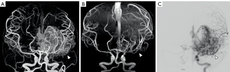
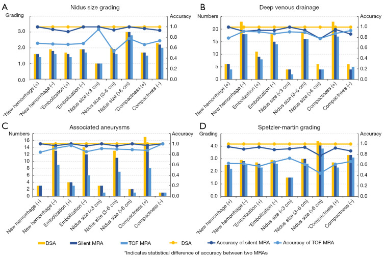
In total, 85 participants (50 males, 35 females; mean age: 33.5±15.2 years) with BAVM who underwent both silent MRA and TOF MRA using a 3 Tesla (3T) magnetic resonance imaging (MRI) system were consecutively recruited from the Capital Medical University Xuanwu Hospital between April 2020 and October 2022 to participate in this cross-sectional retrospective study. The patients were divided into subgroups according to new hemorrhage presentation, embolization, size, and nidus compactness. Image quality scoring on a 4-point scale, and the accuracy of characteristic visulization and Spetzler-Martin grading were compared between the two MRA techniques and each MRA subgroup using the rank-sum Wilcoxon test and Fisher's exact test with digital subtraction angiography (DSA) as the reference standard. A multivariable chi-square test was used to examine the interactions between the grouping factors. A P value <0.05 was considered statistically significant.
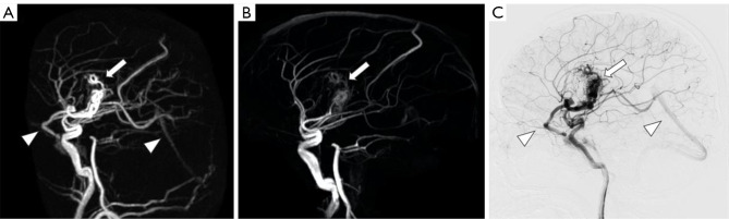
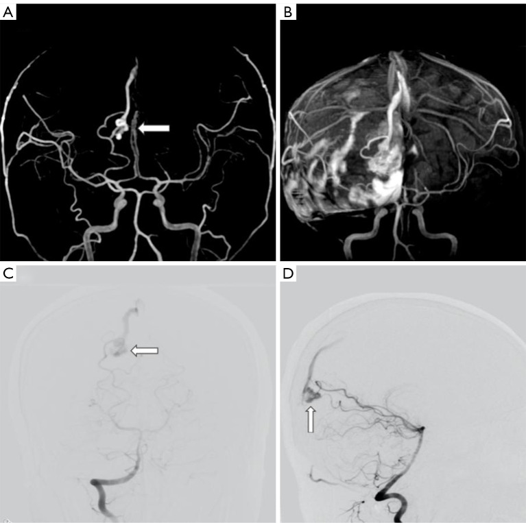
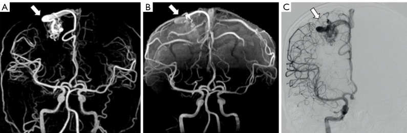
The average image quality scores were significantly higher for silent MRA than those for TOF MRA overall (2.83±0.42 versus 2.46±0.66, P<0.001) and in each subgroup (P<0.05). For silent MRA, the average image quality score for BAVM in each subgroup did not differ significantly (P>0.05). For TOF MRA, the image quality scores for the new hemorrhage, small nidus, and diffuse nidus groups was significantly reduced (P=0.001, <0.001, and 0.037, respectively). The accuracy of silent MRA was significantly better than that of TOF MRA in terms of nidus size and Spetzler-Martin grading (P<0.001), but did not differ significantly in terms of deep venous drainage and associated aneurysm (P=0.402, 0.098, respectively). In relation to silent MRA, the image quality, detection of BAVM characteristics, and grading were similar across the new hemorrhage, embolization, size, and compactness subgroups (P=0.066-0.959). In relation to TOF MRA, the accuracy of nidus size grading was significantly lower in the medium-size subgroup than the small-size subgroup (P<0.001).
Silent MRA performed well in imaging BAVM, and high performance in determining nidus size and Spetzler-Martin grading, but its ability to detect deep venous drainage was limited.
初步的小样本研究表明,在脑动静脉畸形(BAVM)的特征描述方面,静音磁共振血管造影(MRA)优于时间飞跃MRA(TOF MRA),但未研究静音MRA的成像性能是否受BAVM的内在特征或常见临床因素的影响。本研究旨在比较静音MRA和TOF MRA在不同临床情况下对BAVM的可视化和分级情况。
2020年4月至2022年10月期间,从首都医科大学宣武医院连续招募了85例患有BAVM且同时接受了使用3特斯拉(3T)磁共振成像(MRI)系统的静音MRA和TOF MRA检查的参与者(50例男性,35例女性;平均年龄:33.5±15.2岁),参与这项横断面回顾性研究。患者根据新发出血表现、栓塞情况、大小和病灶致密程度分为亚组。采用四点量表对图像质量进行评分,并以数字减影血管造影(DSA)作为参考标准,使用秩和Wilcoxon检验和Fisher精确检验比较两种MRA技术以及每个MRA亚组在特征可视化和Spetzler-Martin分级方面的准确性。采用多变量卡方检验来检验分组因素之间的相互作用。P值<0.05被认为具有统计学意义。
总体而言,静音MRA的平均图像质量评分显著高于TOF MRA(2.83±0.42对2.46±0.66,P<0.001),且在每个亚组中也是如此(P<0.05)。对于静音MRA,各亚组中BAVM的平均图像质量评分差异不显著(P>0.05)。对于TOF MRA,新发出血组、小病灶组和弥漫性病灶组的图像质量评分显著降低(分别为P=0.001、<0.001和0.037)。在病灶大小和Spetzler-Martin分级方面,静音MRA的准确性显著优于TOF MRA(P<0.001),但在深部静脉引流和相关动脉瘤方面差异不显著(分别为P=0.402、0.098)。关于静音MRA,新发出血、栓塞、大小和致密程度亚组在图像质量、BAVM特征检测和分级方面相似(P=0.066 - 0.959)。关于TOF MRA,中等大小亚组在病灶大小分级方面的准确性显著低于小大小亚组(P<0.001)。
静音MRA在BAVM成像方面表现良好,在确定病灶大小和Spetzler-Martin分级方面性能较高,但其检测深部静脉引流的能力有限。