Weil Patrick Philipp, Pembaur Anton, Wirth Beatrice, Oetjen Eda, Büsscher Hannes, Zirngibl Klemens, Czarnetzki Malte, Braun Stella, Cremers Jann-Frederik, Gödde Daniel, Degener Stephan, Postberg Jan
Clinical Molecular Genetics and Epigenetics, Faculty of Health, Centre for Biomedical Education & Research (ZBAF), Witten/Herdecke University, Alfred-Herrhausen-Str. 50, 58448, Witten, Germany.
Centre of Reproductive Medicine and Andrology, University Hospital of Münster, Münster, Germany.
Sci Rep. 2024 Dec 19;14(1):30564. doi: 10.1038/s41598-024-83206-9.
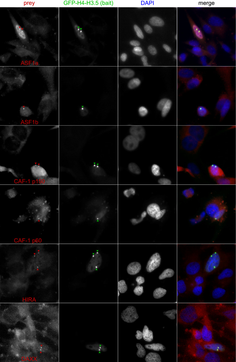
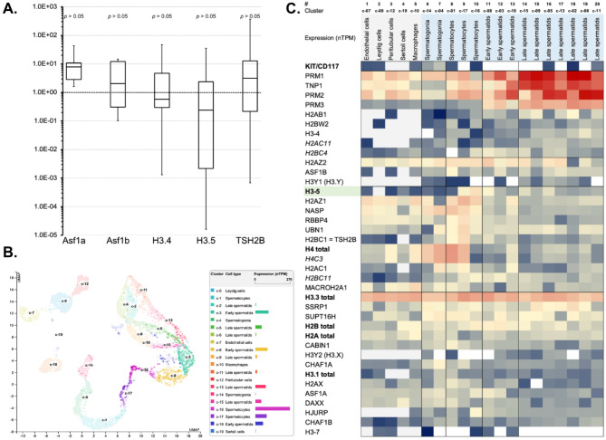
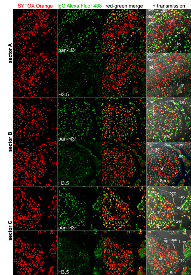
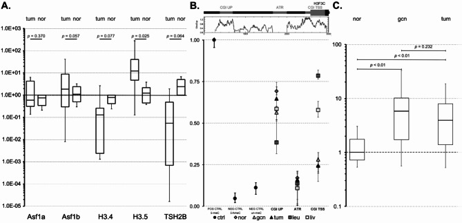
Testicular cell differentiation is a highly regulated process, essential for male reproductive health. The histone variant H3.5 is apparently a critical player in this intricate orchestra of cell types, but its regulation and function remains poorly understood. To elucidate its role, we fractionized testicular cells using c-Kit/CD117 as a separation marker and analyzed H3.5 expression. Further, we investigated the regulation of H3.5 expression using public data repositories. We explored DNA methylation patterns in specific regions of the H3-5 gene and assessed H3-5 copy number gain in seminoma specimens. Additionally, we examined the testicular localization of H3.5 and its histone chaperone interactions to understand its regulation at the protein level. We used qRT-PCR, MeDIP, and qPCR to study H3.5 expression and DNA methylation in various cell types. H3-5 copy number gain was analyzed using qPCR. Protein interactions were investigated through fluorescence-2-hybrid assays in baby hamster kidney cells. H3.5 is primarily enriched in spermatocytes. DNA methylation of a CpG island overlapping the H3-5 promoter appeared to be involved in the tissue-specific regulation of H3.5 expression. Elevated H3.5 expression was observed in seminoma specimens, suggesting a potential link to testicular tumors. H3-5 copy number gain was associated with elevated H3.5 expression in seminoma specimens. Furthermore, we identified physical interactions between H3.5 and histone chaperones Asf1a and Asf1b, HIRA, CAF p150 and DAXX, shedding light on the protein-level regulation of H3.5. These findings provide valuable insights into the molecular mechanisms governing testicular cell differentiation and the potential role of H3.5 in testicular pathologies.
睾丸细胞分化是一个高度受调控的过程,对男性生殖健康至关重要。组蛋白变体H3.5显然是这个复杂细胞类型组合中的关键参与者,但其调控和功能仍知之甚少。为了阐明其作用,我们以c-Kit/CD117作为分离标记对睾丸细胞进行了分级分离,并分析了H3.5的表达。此外,我们利用公共数据储存库研究了H3.5表达的调控。我们探索了H3-5基因特定区域的DNA甲基化模式,并评估了精原细胞瘤标本中H3-5拷贝数增加情况。此外,我们检查了H3.5在睾丸中的定位及其与组蛋白伴侣的相互作用,以了解其在蛋白质水平的调控。我们使用qRT-PCR、MeDIP和qPCR研究了不同细胞类型中H3.5的表达和DNA甲基化。通过qPCR分析H3-5拷贝数增加情况。通过在幼仓鼠肾细胞中进行荧光双杂交试验研究蛋白质相互作用。H3.5主要富集于精母细胞中。与H3-5启动子重叠的一个CpG岛的DNA甲基化似乎参与了H3.5表达的组织特异性调控。在精原细胞瘤标本中观察到H3.5表达升高,提示其与睾丸肿瘤可能存在联系。在精原细胞瘤标本中,H3-5拷贝数增加与H3.5表达升高相关。此外,我们确定了H3.5与组蛋白伴侣Asf1a和Asf1b、HIRA、CAF p150和DAXX之间的物理相互作用,揭示了H3.5在蛋白质水平的调控机制。这些发现为控制睾丸细胞分化的分子机制以及H3.5在睾丸病理中的潜在作用提供了有价值的见解。