Geraghty Terese, Ishihara Shingo, Obeidat Alia M, Adamczyk Natalie S, Hunter Rahel S, Li Jun, Wang Lai, Lee Hoomin, Ko Frank C, Malfait Anne-Marie, Miller Rachel E
Department of Internal Medicine, Division of Rheumatology, Rush University Medical Center, Chicago, IL, USA.
Chicago Center on Musculoskeletal Pain, Chicago, IL, USA.
Arthritis Res Ther. 2024 Dec 20;26(1):224. doi: 10.1186/s13075-024-03457-9.
Osteoarthritis (OA) is a painful degenerative joint disease and a leading source of years lived with disability globally due to inadequate treatment options. Neuroimmune interactions reportedly contribute to OA pain pathogenesis. Notably, in rodents, macrophages in the DRG are associated with onset of persistent OA pain. Our objective was to determine the effects of acute systemic macrophage depletion on pain-related behaviors and joint damage using surgical mouse models in both sexes.
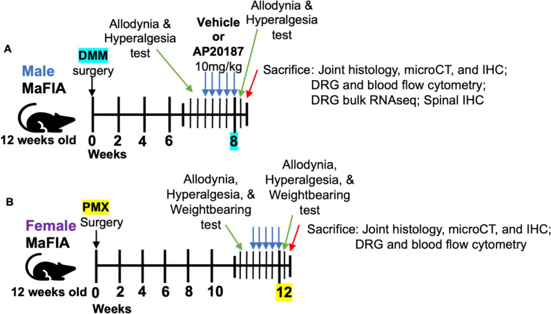
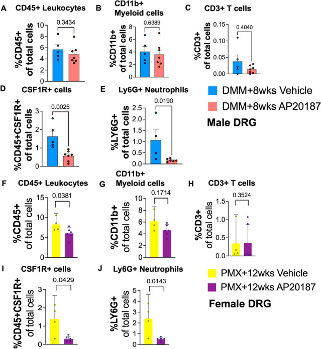
We depleted CSF1R + macrophages by treating male macrophage Fas-induced apoptosis (MaFIA) transgenic mice 8- or 16-weeks post destabilization of the medial meniscus (DMM) with AP20187 or vehicle control (10 mg/kg i.p., 1x/day for 5 days), or treating female MaFIA mice 12 weeks post partial meniscectomy (PMX) with AP20187 or vehicle control. We measured pain-related behaviors 1-3 days before and after depletion, and, 3-4 days after the last injection we examined joint histopathology and performed flow cytometry of the dorsal root ganglia (DRGs). In a separate cohort of male 8-week DMM mice or age-matched naïve vehicle controls, we conducted DRG bulk RNA-sequencing analyses after the 5-day vehicle or AP20187 treatment.
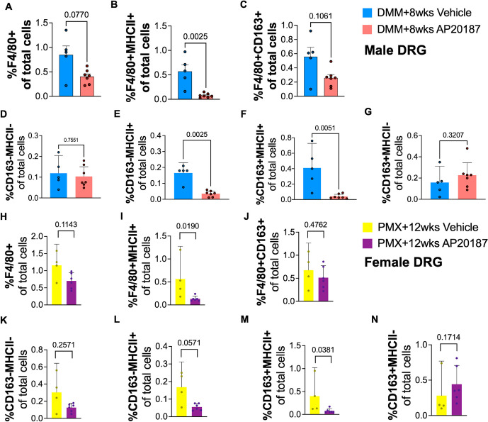
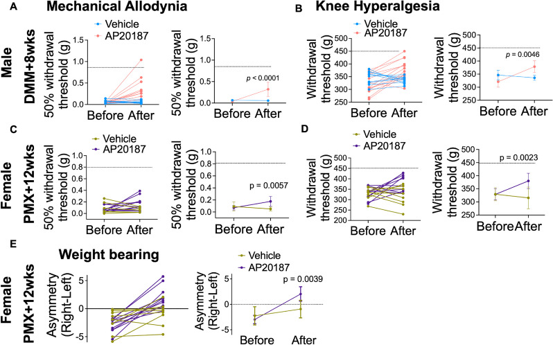
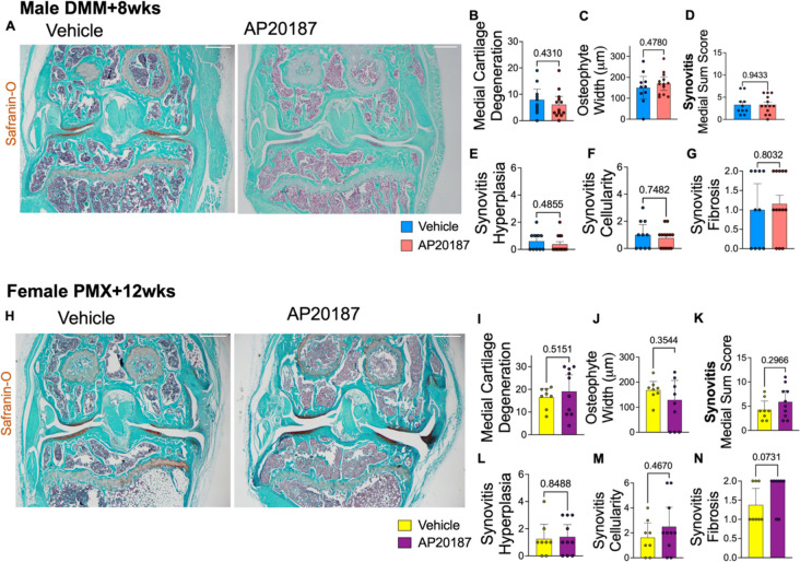
Eight- and 16-weeks post DMM in male mice, AP20187-induced macrophage depletion resulted in attenuated mechanical allodynia and knee hyperalgesia. Female mice showed alleviation of mechanical allodynia, knee hyperalgesia, and weight bearing deficits after macrophage depletion at 12 weeks post PMX. Macrophage depletion did not affect the degree of cartilage degeneration, osteophyte width, or synovitis in either sex. Flow cytometry of the DRG revealed that macrophages and neutrophils were reduced after AP20187 treatment. In addition, in the DRG, only MHCII + M1-like macrophages were significantly decreased, while CD163 + MHCII- M2-like macrophages were not affected in both sexes. DRG bulk RNA-seq revealed that Cxcl10 and Il1b were upregulated with DMM surgery compared to naïve mice, and downregulated in DMM after acute macrophage depletion.
Acute systemic macrophage depletion reduced the levels of pro-inflammatory macrophages in the DRG and alleviated pain-related behaviors in established surgically induced OA in mice of both sexes, without affecting joint damage. Overall, these studies provide insight into immune cell regulation in the DRG during OA.
骨关节炎(OA)是一种疼痛性退行性关节疾病,由于治疗选择有限,是全球残疾生存年数的主要原因。据报道,神经免疫相互作用促成OA疼痛的发病机制。值得注意的是,在啮齿动物中,背根神经节(DRG)中的巨噬细胞与持续性OA疼痛的发作有关。我们的目的是使用手术小鼠模型确定急性全身巨噬细胞耗竭对两性疼痛相关行为和关节损伤的影响。
我们通过在雄性巨噬细胞Fas诱导凋亡(MaFIA)转基因小鼠内侧半月板不稳定(DMM)后8周或16周用AP20187或载体对照(腹腔注射10mg/kg,每天1次,共5天)治疗,或在雌性MaFIA小鼠半月板部分切除术(PMX)后12周用AP20187或载体对照治疗,来耗竭CSF1R +巨噬细胞。我们在耗竭前后1-3天测量疼痛相关行为,在最后一次注射后3-4天检查关节组织病理学并对背根神经节(DRG)进行流式细胞术检测。在另一组雄性8周龄DMM小鼠或年龄匹配的未处理载体对照中,我们在5天载体或AP20187处理后进行DRG批量RNA测序分析。
在雄性小鼠DMM术后8周和16周,AP20187诱导的巨噬细胞耗竭导致机械性异常性疼痛和膝关节痛觉过敏减轻。雌性小鼠在PMX术后12周巨噬细胞耗竭后,机械性异常性疼痛、膝关节痛觉过敏和负重缺陷得到缓解。巨噬细胞耗竭在两性中均未影响软骨退变程度、骨赘宽度或滑膜炎。DRG的流式细胞术显示,AP20187处理后巨噬细胞和中性粒细胞减少。此外,在DRG中,只有MHCII + M1样巨噬细胞显著减少,而CD163 + MHCII- M2样巨噬细胞在两性中均未受影响。DRG批量RNA测序显示,与未处理小鼠相比,DMM手术使Cxcl10和Il1b上调,急性巨噬细胞耗竭后在DMM中下调。
急性全身巨噬细胞耗竭降低了DRG中促炎性巨噬细胞的水平,并减轻了两性小鼠既定手术诱导的OA中的疼痛相关行为,而不影响关节损伤。总体而言,这些研究为OA期间DRG中的免疫细胞调节提供了见解。