Enninful Archibald, Zhang Zhaojun, Klymyshyn Dmytro, Zong Hailing, Bai Zhiliang, Farzad Negin, Su Graham, Baysoy Alev, Nam Jungmin, Yang Mingyu, Lu Yao, Zhang Nancy R, Braubach Oliver, Xu Mina L, Ma Zongming, Fan Rong
Department of Biomedical Engineering, Yale University, New Haven, CT, 06520, USA.
Department of Statistics and Data Science, The Wharton School, University of Pennsylvania, Philadelphia, PA, USA.
Res Sq. 2024 Dec 11:rs.3.rs-5398491. doi: 10.21203/rs.3.rs-5398491/v1.
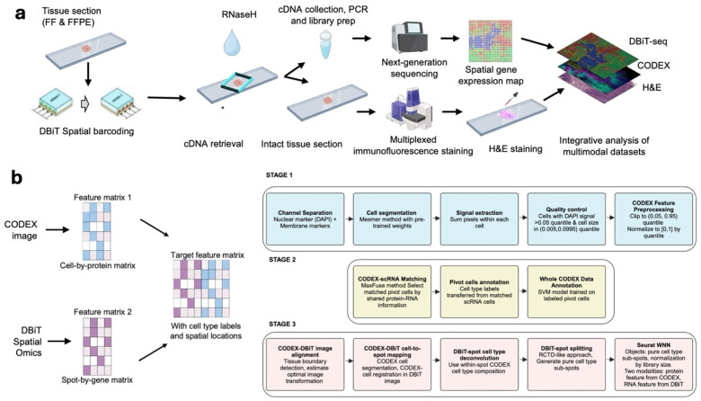
Spatially mapping the transcriptome and proteome in the same tissue section can significantly advance our understanding of heterogeneous cellular processes and connect cell type to function. Here, we present Deterministic Barcoding in Tissue sequencing plus (DBiTplus), an integrative multi-modality spatial omics approach that combines sequencing-based spatial transcriptomics and image-based spatial protein profiling on the same tissue section to enable both single-cell resolution cell typing and genome-scale interrogation of biological pathways. DBiTplus begins with reverse transcription for cDNA synthesis, microfluidic delivery of DNA oligos for spatial barcoding, retrieval of barcoded cDNA using RNaseH, an enzyme that selectively degrades RNA in an RNA-DNA hybrid, preserving the intact tissue section for high-plex protein imaging with CODEX. We developed computational pipelines to register data from two distinct modalities. Performing both DBiT-seq and CODEX on the same tissue slide enables accurate cell typing in each spatial transcriptome spot and subsequently image-guided decomposition to generate single-cell resolved spatial transcriptome atlases. DBiTplus was applied to mouse embryos with limited protein markers but still demonstrated excellent integration for single-cell transcriptome decomposition, to normal human lymph nodes with high-plex protein profiling to yield a single-cell spatial transcriptome map, and to human lymphoma FFPE tissue to explore the mechanisms of lymphomagenesis and progression. DBiTplusCODEX is a unified workflow including integrative experimental procedure and computational innovation for spatially resolved single-cell atlasing and exploration of biological pathways cell-by-cell at genome-scale.
在同一组织切片上对转录组和蛋白质组进行空间映射,可以显著推进我们对异质细胞过程的理解,并将细胞类型与功能联系起来。在这里,我们展示了组织测序增强型确定性条形码技术(DBiTplus),这是一种整合的多模态空间组学方法,它将基于测序的空间转录组学和基于图像的空间蛋白质分析结合在同一组织切片上,以实现单细胞分辨率的细胞分型和对生物途径的全基因组规模研究。DBiTplus首先进行逆转录以合成cDNA,通过微流控技术递送DNA寡核苷酸进行空间条形码标记,使用RNaseH检索条形码化的cDNA,RNaseH是一种能选择性降解RNA-DNA杂交体中RNA的酶,从而保留完整的组织切片用于通过CODEX进行高多重蛋白质成像。我们开发了计算管道来整合来自两种不同模态的数据。在同一张组织载玻片上同时进行DBiT-seq和CODEX,能够在每个空间转录组斑点中进行准确的细胞分型,随后通过图像引导分解生成单细胞分辨率的空间转录组图谱。DBiTplus被应用于蛋白质标记有限的小鼠胚胎,但仍展示了用于单细胞转录组分解的出色整合能力,应用于具有高多重蛋白质分析的正常人淋巴结以生成单细胞空间转录组图谱,还应用于人类淋巴瘤FFPE组织以探索淋巴瘤发生和进展的机制。DBiTplus-CODEX是一个统一的工作流程,包括整合的实验程序和计算创新,用于在全基因组规模上逐个细胞地进行空间分辨的单细胞图谱绘制和生物途径探索。