• 文献检索
  • 文档翻译
  • 深度研究
  • 学术资讯
  • Suppr Zotero 插件Zotero 插件
  • 邀请有礼
  • 套餐&价格
  • 历史记录
应用&插件
Suppr Zotero 插件Zotero 插件浏览器插件Mac 客户端Windows 客户端微信小程序
定价
高级版会员购买积分包购买API积分包
服务
文献检索文档翻译深度研究API 文档MCP 服务
关于我们
关于 Suppr公司介绍联系我们用户协议隐私条款
关注我们

Suppr 超能文献

核心技术专利:CN118964589B侵权必究
粤ICP备2023148730 号-1Suppr @ 2026

文献检索

告别复杂PubMed语法,用中文像聊天一样搜索,搜遍4000万医学文献。AI智能推荐,让科研检索更轻松。

立即免费搜索

文件翻译

保留排版,准确专业,支持PDF/Word/PPT等文件格式,支持 12+语言互译。

免费翻译文档

深度研究

AI帮你快速写综述,25分钟生成高质量综述,智能提取关键信息,辅助科研写作。

立即免费体验

结节周围透亮征作为单侧股骨头骨骺滑脱病例中对侧髋关节滑脱的一个重要预测因素。

The Peritubercle Lucency Sign as a Considerable Predictive Factor for Contralateral Hip Slippage in Unilateral Slipped Capital Femoral Epiphysis Cases.

作者信息

Neto Anastácio Kotzias, Martinelli Renan Vinicius Romano, Gressler Marthina Alice, Oliveira Marco Aurélio de

机构信息

Departamento de Ortopedia Pediátrica, Hospital Infantil Joana de Gusmão, Florianópolis, SC, Brasil.

出版信息

Rev Bras Ortop (Sao Paulo). 2024 Dec 21;59(6):e922-e935. doi: 10.1055/s-0044-1790578. eCollection 2024 Dec.

DOI:10.1055/s-0044-1790578
PMID:39711631
原文链接:https://pmc.ncbi.nlm.nih.gov/articles/PMC11663065/
Abstract

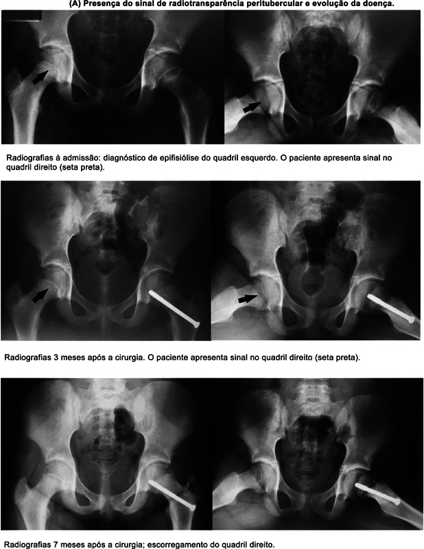
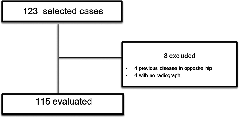
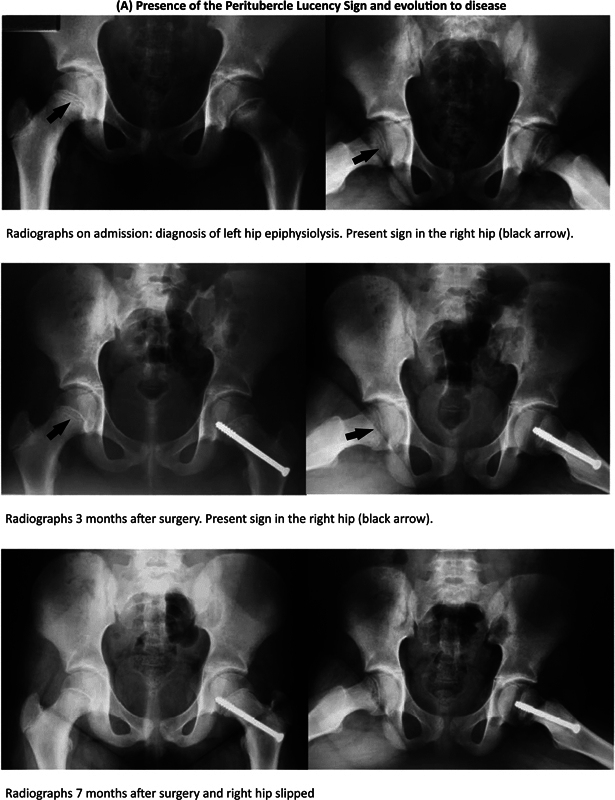
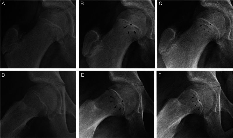
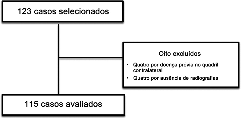
To determine whether the radiographic parameter at the epiphyseal tubercle region (peritubercle lucency sign) on the unaffected side can predict slipped capital femoral epiphysis (SCFE).  We retrospectively reviewed patients who received an initial diagnosis of unilateral SCFE between 1995 and 2020 at a pediatric hospital in a Brazilian state's capital. The patients were monitored for at least 18 months. Two reviewers independently and blindly assessed the radiographs for the presence or absence of the sign. Disagreements were resolved by a third senior reviewer.  Out of the 115 cases reviewed, the peritubercle lucency sign was observed in 21 of the 30 patients who developed the disease in the contralateral hip. The sign was observed on an average of 21 days after the diagnosis on the initial side, and approximately 301 days prior to the condition affecting the contralateral hip. It was present in 95% and 85% of the cases on the lateral (frog-leg) and anteroposterior (AP) views, respectively. Interobserver reliability was measured using the Kappa test (k = 0.0801). There was a significant relationship between the presence of the sign and SCFE (  < 0.001).  We propose that the peritubercle lucency sign can be used as a supplementary tool in early diagnosis, for it is beneficial in the therapeutic planning.  Level III - Diagnostic Study In Nonconsecutive Patients (Without Consistently Applied 'Gold Standard' As Reference).

摘要

为了确定未受影响侧骨骺结节区域的影像学参数(结节周围透亮征)是否能够预测股骨头骨骺滑脱(SCFE)。

我们回顾性分析了1995年至2020年期间在巴西某州首府一家儿科医院首次诊断为单侧SCFE的患者。对这些患者进行了至少18个月的监测。两名评估人员独立且盲法评估X线片上是否存在该征象。分歧由第三位资深评估人员解决。

在115例回顾病例中,对侧髋关节发病的30例患者中有21例观察到结节周围透亮征。该征象在初始侧诊断后平均21天出现,在对侧髋关节发病前约301天出现。分别在95%和85%的病例中,该征象在蛙式侧位和前后位片上可见。采用Kappa检验测量观察者间信度(k = 0.0801)。该征象的存在与SCFE之间存在显著相关性(P < 0.001)。

我们建议结节周围透亮征可作为早期诊断的辅助工具,因为它有助于治疗方案的制定。

三级——非连续患者的诊断性研究(未始终采用“金标准”作为参考)

https://cdn.ncbi.nlm.nih.gov/pmc/blobs/015a/11663065/1a837c5ea3b0/10-1055-s-0044-1790578-i2400118pt-3.jpg
https://cdn.ncbi.nlm.nih.gov/pmc/blobs/015a/11663065/ee79e35ede51/10-1055-s-0044-1790578-i2400118en-1.jpg
https://cdn.ncbi.nlm.nih.gov/pmc/blobs/015a/11663065/8240545d7dc1/10-1055-s-0044-1790578-i2400118en-2.jpg
https://cdn.ncbi.nlm.nih.gov/pmc/blobs/015a/11663065/9888c376846f/10-1055-s-0044-1790578-i2400118en-3.jpg
https://cdn.ncbi.nlm.nih.gov/pmc/blobs/015a/11663065/9f182e30da40/10-1055-s-0044-1790578-i2400118pt-1.jpg
https://cdn.ncbi.nlm.nih.gov/pmc/blobs/015a/11663065/036cc5da88e7/10-1055-s-0044-1790578-i2400118pt-2.jpg
https://cdn.ncbi.nlm.nih.gov/pmc/blobs/015a/11663065/1a837c5ea3b0/10-1055-s-0044-1790578-i2400118pt-3.jpg

相似文献

1
The Peritubercle Lucency Sign as a Considerable Predictive Factor for Contralateral Hip Slippage in Unilateral Slipped Capital Femoral Epiphysis Cases.结节周围透亮征作为单侧股骨头骨骺滑脱病例中对侧髋关节滑脱的一个重要预测因素。
Rev Bras Ortop (Sao Paulo). 2024 Dec 21;59(6):e922-e935. doi: 10.1055/s-0044-1790578. eCollection 2024 Dec.
2
What Is the Accuracy and Reliability of the Peritubercle Lucency Sign on Radiographs for Early Diagnosis of Slipped Capital Femoral Epiphysis Compared With MRI as the Gold Standard?X 线片上的干骺端透亮线征象对早期诊断股骨颈骨骺滑脱的准确性和可靠性与 MRI 作为金标准相比如何?
Clin Orthop Relat Res. 2020 May;478(5):1049-1059. doi: 10.1097/CORR.0000000000001136.
3
The Peritubercle Lucency Sign is a Common and Early Radiographic Finding in Slipped Capital Femoral Epiphysis.结节周围透亮征是股骨头骨骺滑脱常见的早期影像学表现。
J Pediatr Orthop. 2018 Aug;38(7):e371-e376. doi: 10.1097/BPO.0000000000001198.
4
Intraobserver and Interobserver Reliability of the Peritubercle Lucency Sign in Slipped Capital Femoral Epiphysis.滑向性股骨骨骺滑脱症中干骺端透亮线征象的观察者内和观察者间可靠性。
J Pediatr Orthop. 2021 Mar 1;41(3):159-163. doi: 10.1097/BPO.0000000000001733.
5
Morphologic Features of the Contralateral Femur in Patients With Unilateral Slipped Capital Femoral Epiphysis Resembles Mild Slip Deformity: A Matched Cohort Study.单侧股骨头骨骺滑脱患者对侧股骨的形态特征类似于轻度滑脱畸形:一项配对队列研究。
Clin Orthop Relat Res. 2018 Apr;476(4):890-899. doi: 10.1007/s11999.0000000000000127.
6
The S Sign: A New Radiographic Tool to Aid in the Diagnosis of Slipped Capital Femoral Epiphysis.S征:一种有助于诊断股骨头骨骺滑脱的新型影像学工具。
J Emerg Med. 2018 Jun;54(6):835-843. doi: 10.1016/j.jemermed.2018.01.023. Epub 2018 Mar 15.
7
How Common Is Femoral Retroversion and How Is it Affected by Different Measurement Methods in Unilateral Slipped Capital Femoral Epiphysis?股骨后旋在单侧股骨头骨骺滑脱中有多常见,不同的测量方法对其有何影响?
Clin Orthop Relat Res. 2021 May 1;479(5):947-959. doi: 10.1097/CORR.0000000000001611.
8
Radiology of adolescent slipped capital femoral epiphysis: measurement of epiphyseal angles and diagnosis.青少年股骨头骨骺滑脱的放射学:骨骺角的测量与诊断
Oper Orthop Traumatol. 2007 Oct;19(4):329-44. doi: 10.1007/s00064-007-1214-6.
9
Capital Femoral Epiphyseal Cupping and Extension May Be Protective in Slipped Capital Femoral Epiphysis: A Dual-center Matching Cohort Study.股骨近端骨骺杯状化和延长可能对股骨头骨骺滑脱具有保护作用:一项双中心匹配队列研究。
J Pediatr Orthop. 2020 Aug;40(7):334-339. doi: 10.1097/BPO.0000000000001528.
10
What Is the Prevalence of Cam Deformity After Prophylactic Pinning of the Contralateral Asymptomatic Hip in Unilateral Slipped Capital Femoral Epiphysis? A 10-year Minimum Followup Study.预防性固定对侧无症状髋关节后凸轮畸形的发生率是多少?一项 10 年的最低随访研究。
Clin Orthop Relat Res. 2019 May;477(5):1111-1122. doi: 10.1097/CORR.0000000000000592.

本文引用的文献

1
Intraobserver and Interobserver Reliability of the Peritubercle Lucency Sign in Slipped Capital Femoral Epiphysis.滑向性股骨骨骺滑脱症中干骺端透亮线征象的观察者内和观察者间可靠性。
J Pediatr Orthop. 2021 Mar 1;41(3):159-163. doi: 10.1097/BPO.0000000000001733.
2
CORR Insights®: What is the Accuracy and Reliability of the Peritubercle Lucency Sign on Radiographs for Early Diagnosis of Slipped Capital Femoral Epiphysis Compared With MRI as the Gold Standard?CORR见解®:与作为金标准的MRI相比,X线片上的骨骺周围透亮征在早期诊断股骨头骨骺滑脱中的准确性和可靠性如何?
Clin Orthop Relat Res. 2020 May;478(5):1060-1061. doi: 10.1097/CORR.0000000000001232.
3
What Is the Accuracy and Reliability of the Peritubercle Lucency Sign on Radiographs for Early Diagnosis of Slipped Capital Femoral Epiphysis Compared With MRI as the Gold Standard?
X 线片上的干骺端透亮线征象对早期诊断股骨颈骨骺滑脱的准确性和可靠性与 MRI 作为金标准相比如何?
Clin Orthop Relat Res. 2020 May;478(5):1049-1059. doi: 10.1097/CORR.0000000000001136.
4
Predicting Risk of Contralateral Slip in Unilateral Slipped Capital Femoral Epiphysis: Posterior Epiphyseal Tilt Increases and Superior Epiphyseal Extension Reduces Risk.预测单侧股骨头骨骺滑脱对侧滑移的风险:后骨骺倾斜增加和上骨骺延伸减少风险。
J Bone Joint Surg Am. 2019 Feb 6;101(3):209-217. doi: 10.2106/JBJS.18.00440.
5
Age- and Sex-Specific Morphologic Variations of Capital Femoral Epiphysis Growth in Children and Adolescents Without Hip Disorders.无髋关节疾病的儿童和青少年股骨头骨骺生长的年龄和性别特异性形态学变异
Orthop J Sports Med. 2018 Jun 25;6(6):2325967118781579. doi: 10.1177/2325967118781579. eCollection 2018 Jun.
6
The Peritubercle Lucency Sign is a Common and Early Radiographic Finding in Slipped Capital Femoral Epiphysis.结节周围透亮征是股骨头骨骺滑脱常见的早期影像学表现。
J Pediatr Orthop. 2018 Aug;38(7):e371-e376. doi: 10.1097/BPO.0000000000001198.
7
Radiographic evaluation in epiphysiolysis: possible predictors of bilaterality?骨骺分离的影像学评估:双侧性的可能预测因素?
Acta Ortop Bras. 2012;20(4):203-6. doi: 10.1590/S1413-78522012000400001.
8
An anatomic study of the epiphyseal tubercle and its importance in the pathogenesis of slipped capital femoral epiphysis.骺结节的解剖学研究及其在股骨头骨骺滑脱发病机制中的重要性。
J Bone Joint Surg Am. 2013 Mar 20;95(6):e341-8. doi: 10.2106/JBJS.L.00474.
9
Femoral morphology differs between deficient and excessive acetabular coverage.髋臼覆盖不足与过度时,股骨形态会有所不同。
Clin Orthop Relat Res. 2008 Apr;466(4):782-90. doi: 10.1007/s11999-008-0141-7. Epub 2008 Feb 21.
10
Does the upper femoral epiphysis slip or rotate?股骨近端骨骺会发生滑脱还是旋转?
J Bone Joint Surg Br. 2007 Oct;89(10):1402-6. doi: 10.1302/0301-620X.89B10.19563.