• 文献检索
  • 文档翻译
  • 深度研究
  • 学术资讯
  • Suppr Zotero 插件Zotero 插件
  • 邀请有礼
  • 套餐&价格
  • 历史记录
应用&插件
Suppr Zotero 插件Zotero 插件浏览器插件Mac 客户端Windows 客户端微信小程序
定价
高级版会员购买积分包购买API积分包
服务
文献检索文档翻译深度研究API 文档MCP 服务
关于我们
关于 Suppr公司介绍联系我们用户协议隐私条款
关注我们

Suppr 超能文献

核心技术专利:CN118964589B侵权必究
粤ICP备2023148730 号-1Suppr @ 2026

文献检索

告别复杂PubMed语法,用中文像聊天一样搜索,搜遍4000万医学文献。AI智能推荐,让科研检索更轻松。

立即免费搜索

文件翻译

保留排版,准确专业,支持PDF/Word/PPT等文件格式,支持 12+语言互译。

免费翻译文档

深度研究

AI帮你快速写综述,25分钟生成高质量综述,智能提取关键信息,辅助科研写作。

立即免费体验

上消化道疾病的图像增强内镜检查:聚焦于纹理和颜色增强成像以及红色双色成像。

Image-enhanced endoscopy in upper gastrointestinal disease: focusing on texture and color enhancement imaging and red dichromatic imaging.

作者信息

Park Jae Yong

机构信息

Division of Gastroenterology, Department of Internal Medicine, Chung-Ang University College of Medicine, Seoul, Korea.

出版信息

Clin Endosc. 2025 Mar;58(2):163-180. doi: 10.5946/ce.2024.159. Epub 2024 Nov 6.

DOI:10.5946/ce.2024.159
PMID:39722144
原文链接:https://pmc.ncbi.nlm.nih.gov/articles/PMC11983135/
Abstract

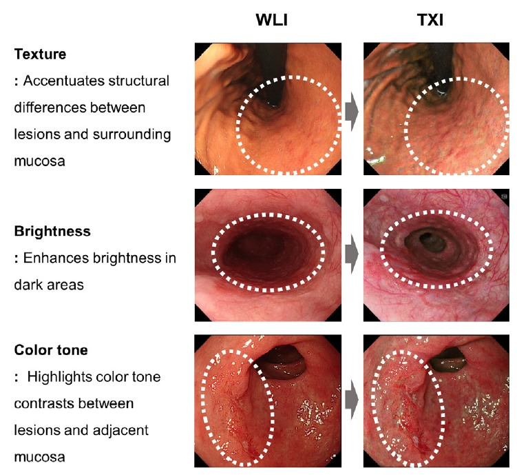
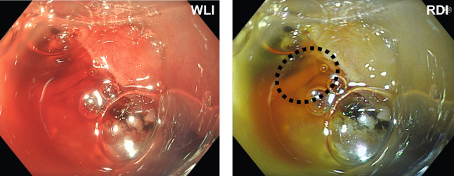
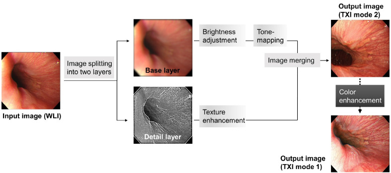
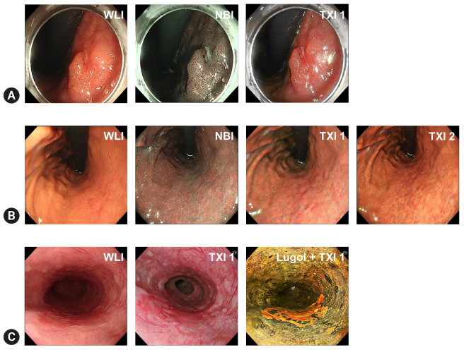
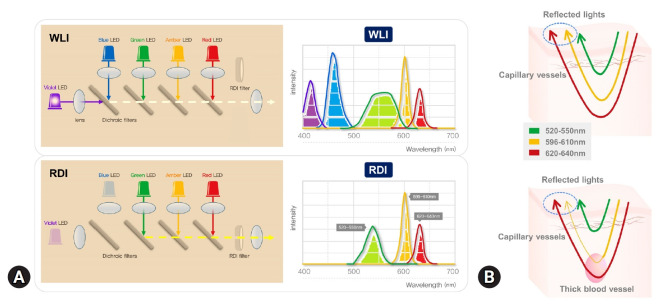
Endoscopic examination plays a crucial role in the diagnosis of upper gastrointestinal (UGI) tract diseases. Despite advancements in endoscopic imaging, the detection of subtle early cancers and premalignant lesions using white-light imaging alone remains challenging. This review discusses two novel image-enhanced endoscopy (IEE) techniques-texture and color enhancement imaging (TXI) and red dichromatic imaging (RDI)-and their potential applications in UGI diseases. TXI enhances texture, brightness, and color tone, which improves the visibility of mucosal irregularities and facilitates earlier detection of neoplastic lesions. Studies have suggested that TXI enhances the color differences between lesions and the surrounding mucosa and improves the visibility of the lesion. TXI aids in the diagnosis of various UGI diseases, including early gastric cancer, esophageal cancer, premalignant conditions such as atrophic gastritis and Barrett's esophagus, and duodenal tumors. RDI utilizes specific wavelengths to enhance the visualization of deep blood vessels or bleeding points, aiding in the rapid and accurate identification of bleeding sources during endoscopic procedures. Although promising, TXI and RDI require further large-scale studies across diverse populations to establish their clinical utility, diagnostic performance, and cost-effectiveness before integration into the guidelines. Standardized training is also required for effective utilization. Overall, these IEE techniques has the potential to improve the diagnosis and management of UGI.

摘要

内镜检查在上消化道(UGI)疾病的诊断中起着至关重要的作用。尽管内镜成像技术不断进步,但仅使用白光成像检测细微的早期癌症和癌前病变仍然具有挑战性。本文综述了两种新型的图像增强内镜检查(IEE)技术——纹理和色彩增强成像(TXI)以及红二色成像(RDI)——及其在上消化道疾病中的潜在应用。TXI可增强纹理、亮度和色调,从而提高黏膜不规则处的可见性,并有助于更早地发现肿瘤性病变。研究表明,TXI可增强病变与周围黏膜之间的颜色差异,提高病变的可见性。TXI有助于诊断各种上消化道疾病,包括早期胃癌、食管癌、萎缩性胃炎和巴雷特食管等癌前病变以及十二指肠肿瘤。RDI利用特定波长来增强深部血管或出血点的可视化,有助于在内镜检查过程中快速准确地识别出血源。尽管前景广阔,但TXI和RDI在纳入指南之前,需要在不同人群中进行进一步的大规模研究,以确定其临床实用性、诊断性能和成本效益。有效利用这些技术还需要标准化培训。总体而言,这些IEE技术有潜力改善上消化道疾病的诊断和管理。

https://cdn.ncbi.nlm.nih.gov/pmc/blobs/e002/11983135/b5c1ddbfafb9/ce-2024-159f5.jpg
https://cdn.ncbi.nlm.nih.gov/pmc/blobs/e002/11983135/803886961da5/ce-2024-159f1.jpg
https://cdn.ncbi.nlm.nih.gov/pmc/blobs/e002/11983135/df5c6cadda2c/ce-2024-159f2.jpg
https://cdn.ncbi.nlm.nih.gov/pmc/blobs/e002/11983135/2e2af80f9aae/ce-2024-159f3.jpg
https://cdn.ncbi.nlm.nih.gov/pmc/blobs/e002/11983135/90a0e4d3d046/ce-2024-159f4.jpg
https://cdn.ncbi.nlm.nih.gov/pmc/blobs/e002/11983135/b5c1ddbfafb9/ce-2024-159f5.jpg

相似文献

1
Image-enhanced endoscopy in upper gastrointestinal disease: focusing on texture and color enhancement imaging and red dichromatic imaging.上消化道疾病的图像增强内镜检查:聚焦于纹理和颜色增强成像以及红色双色成像。
Clin Endosc. 2025 Mar;58(2):163-180. doi: 10.5946/ce.2024.159. Epub 2024 Nov 6.
2
Using texture and colour enhancement imaging to evaluate gastrointestinal diseases in clinical practice: a review.利用纹理和色彩增强成像技术评估临床胃肠道疾病:综述。
Ann Med. 2022 Dec;54(1):3315-3332. doi: 10.1080/07853890.2022.2147992.
3
Third-Generation High-Vision Ultrathin Endoscopy Using Texture and Color Enhancement Imaging and Narrow-Band Imaging to Evaluate Barrett's Esophagus.使用纹理和颜色增强成像以及窄带成像的第三代高清超薄内镜检查法评估巴雷特食管
Diagnostics (Basel). 2022 Dec 13;12(12):3149. doi: 10.3390/diagnostics12123149.
4
Recent advancements in image-enhanced endoscopy in the pancreatobiliary field.胰胆领域图像增强内镜检查的最新进展。
DEN Open. 2024 May 14;5(1):e382. doi: 10.1002/deo2.382. eCollection 2025 Apr.
5
Texture and Color Enhancement Imaging Increases Color Changes and Improves Visibility for Squamous Cell Carcinoma Suspicious Lesions in the Pharynx and Esophagus.纹理与颜色增强成像增加了颜色变化并提高了咽部和食管鳞状细胞癌可疑病变的可视性。
Diagnostics (Basel). 2021 Oct 23;11(11):1971. doi: 10.3390/diagnostics11111971.
6
Emerging texture and color enhancement imaging in early gastric cancer.早期胃癌的新兴纹理和色彩增强成像技术。
Dig Endosc. 2022 May;34(4):714-720. doi: 10.1111/den.14182. Epub 2021 Dec 1.
7
Assessing the effectiveness of texture and color enhancement imaging versus white-light endoscopy in detecting gastrointestinal lesions: A systematic review and meta-analysis.评估纹理和颜色增强成像与白光内镜检查在检测胃肠道病变中的有效性:一项系统评价和荟萃分析。
DEN Open. 2025 Apr 30;6(1):e70128. doi: 10.1002/deo2.70128. eCollection 2026 Apr.
8
Comparison of Texture and Color Enhancement Imaging with White Light Imaging in 52 Patients with Short-Segment Barrett's Esophagus.52 例短节段 Barrett 食管患者的纹理及色彩增强成像与白光成像的对比。
Med Sci Monit. 2023 Jun 13;29:e940249. doi: 10.12659/MSM.940249.
9
TXI: Texture and Color Enhancement Imaging for Endoscopic Image Enhancement.TXI:用于内窥镜图像增强的纹理和颜色增强成像。
J Healthc Eng. 2021 Apr 7;2021:5518948. doi: 10.1155/2021/5518948. eCollection 2021.
10
Endoscopic submucosal dissection and tunneling procedures using novel image-enhanced technique.使用新型图像增强技术的内镜黏膜下剥离术和隧道技术。
VideoGIE. 2022 Jan 5;7(4):158-163. doi: 10.1016/j.vgie.2021.11.005. eCollection 2022 Apr.

引用本文的文献

1
Navigating the Treatment Landscape for Widespread Superficial Esophageal Squamous Cell Neoplasia.应对广泛浅表性食管鳞状细胞瘤的治疗局面
Gut Liver. 2025 May 15;19(3):305-306. doi: 10.5009/gnl250084.
2
Classification of image-enhanced endoscopy in colon tumors.结肠肿瘤的图像增强内镜检查分类
Clin Endosc. 2025 May;58(3):337-351. doi: 10.5946/ce.2024.263. Epub 2025 May 8.

本文引用的文献

1
Predicting Helicobacter pylori infection from endoscopic features.从内镜特征预测幽门螺杆菌感染。
Korean J Intern Med. 2024 May;39(3):439-447. doi: 10.3904/kjim.2023.300. Epub 2024 Apr 30.
2
Endoscopic submucosal dissection for superficial esophageal cancer with ulcer scarring using a combination of pocket creation, gel immersion, and red dichromatic imaging.采用建立袋状结构、凝胶浸润和红色双色成像相结合的方法,对伴有溃疡瘢痕的浅表食管癌进行内镜黏膜下剥离术。
Endoscopy. 2024 Dec;56(S 01):E87-E88. doi: 10.1055/a-2234-8435. Epub 2024 Jan 30.
3
Advanced endoscopic imaging for detection of Barrett's esophagus.
用于检测巴雷特食管的先进内镜成像技术。
Clin Endosc. 2024 Jan;57(1):1-10. doi: 10.5946/ce.2023.031. Epub 2024 Jan 5.
4
Texture and color enhancement imaging improves the visibility of gastric neoplasms: clinical trial with image catalogue assessment using conventional and newly developed endoscopes.纹理和色彩增强成像提高胃肿瘤的可视性:使用传统和新开发的内镜进行图像目录评估的临床试验。
BMC Gastroenterol. 2023 Nov 13;23(1):389. doi: 10.1186/s12876-023-03030-9.
5
Red Dichromatic Imaging Improves the Recognition of Bleeding Points During Endoscopic Submucosal Dissection.红色双色色差成像提高内镜黏膜下剥离术中出血点的识别率。
Dig Dis Sci. 2024 Jan;69(1):216-227. doi: 10.1007/s10620-023-08145-3. Epub 2023 Nov 13.
6
Diagnosis and management of Barrett esophagus: European Society of Gastrointestinal Endoscopy (ESGE) Guideline.巴雷特食管的诊断和管理:欧洲胃肠道内镜学会(ESGE)指南。
Endoscopy. 2023 Dec;55(12):1124-1146. doi: 10.1055/a-2176-2440. Epub 2023 Oct 9.
7
Endoscopic Submucosal Dissection for Esophageal Squamous Cell Carcinoma With Varices Using Red Dichromatic Imaging.使用红色双色成像技术对伴有静脉曲张的食管鳞状细胞癌进行内镜黏膜下剥离术。
Clin Gastroenterol Hepatol. 2024 Feb;22(2):A31-A32. doi: 10.1016/j.cgh.2023.08.036. Epub 2023 Sep 14.
8
Image enhanced colonoscopy: updates and prospects-a review.图像增强结肠镜检查:最新进展与前景——综述
Transl Gastroenterol Hepatol. 2023 Jul 25;8:26. doi: 10.21037/tgh-23-17. eCollection 2023.
9
Higher detectability of gastric cancer after eradication in texture and color enhancement imaging mode 2 in screening endoscopy.在筛查内镜检查的纹理和颜色增强成像模式2下,根除幽门螺杆菌后胃癌的可检测性更高。 (注:原文中未提及“根除幽门螺杆菌”,根据语境推测可能是相关背景信息遗漏,若实际原文无误,可忽略此注释内容)
DEN Open. 2023 Jul 30;4(1):e279. doi: 10.1002/deo2.279. eCollection 2024 Apr.
10
Usefulness of texture- and color-enhancement imaging for identifying the bleeding point in a patient with post-endoscopic sphincterotomy bleeding.纹理和颜色增强成像在识别内镜下括约肌切开术后出血患者出血点中的应用价值。
VideoGIE. 2023 Apr 14;8(7):269-271. doi: 10.1016/j.vgie.2023.02.013. eCollection 2023 Jul.