Petzer Melissa, Fobian Seth-Frerich, Gulumian Mary, Steenkamp Vanessa, Cordier Werner
Department of Pharmacology, Faculty of Health Sciences, University of Pretoria, Pretoria, South Africa.
Molecular Medicine and Haematology, School of Pathology, University of Witwatersrand, Johannesburg, South Africa.
Pharmacol Res Perspect. 2025 Feb;13(1):e70051. doi: 10.1002/prp2.70051.
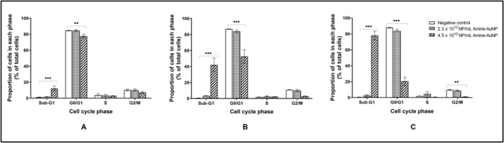
Gold nanoparticles (AuNPs) present with unique physicochemical features and potential for functionalization as anticancer agents. Three-dimensional spheroid models can be used to afford greater tissue representation due to their heterogeneous phenotype and complex molecular architecture. This study developed an A549 alveolar carcinoma spheroid model for cytotoxicity assessment and mechanistic evaluation of functionalized AuNPs. A549 spheroids were generated using an agarose micro-mold and were characterized (morphology, acid phosphatase activity, protein content) over 21 culturing days. The 72-h cytotoxicity of carboxyl-polyethylene glycol- (PCOOH-) and amine-polyethylene glycol- (PNH-) functionalized AuNPs against Day 7 spheroids was assessed by determining spheroid morphology, acid phosphatase activity, protein content, caspase-3/7 activity, and cell cycle kinetics. Spheroids remained stable over the experimental period. Although the A549 spheroids' volume increased while remaining viable over the culturing period, structural integrity decreased from Day 14 onwards. The PCOOH-AuNPs lacked cytotoxicity at a maximum concentration of 1.2 × 10 nanoparticles/mL with no prominent alteration to the cellular processes investigated, while the PNH-AuNPs (at a maximum of 4.5 × 10 nanoparticles/mL) displayed dose- and time-dependent cytotoxicity with associated loss of spheroid compactness, debris formation, DNA fragmentation, and a 75% reduction in acid phosphatase activity. Differentiation between cytotoxic and non-cytotoxic AuNPs was achieved, with preliminary elucidation of cytotoxicity endpoints. The PNH-AuNPs promote cytotoxicity by modulating cellular kinetics while destabilizing the spheroid ultrastructure. The model serves as a proficient platform for more in-depth elucidation of NP cytotoxicity at the preclinical investigation phase.
金纳米颗粒(AuNPs)具有独特的物理化学特性以及作为抗癌剂进行功能化修饰的潜力。三维球体模型由于其异质表型和复杂的分子结构,可用于提供更接近组织的表现形式。本研究开发了一种用于功能化AuNPs细胞毒性评估和机制评价的A549肺泡癌球体模型。使用琼脂糖微模具生成A549球体,并在21天的培养过程中对其进行表征(形态、酸性磷酸酶活性、蛋白质含量)。通过测定球体形态、酸性磷酸酶活性、蛋白质含量、半胱天冬酶-3/7活性和细胞周期动力学,评估羧基聚乙二醇(PCOOH-)和胺基聚乙二醇(PNH-)功能化AuNPs对第7天球体的72小时细胞毒性。在实验期间,球体保持稳定。尽管A549球体的体积在培养期间增加且仍保持活力,但其结构完整性从第14天起开始下降。PCOOH-AuNPs在最大浓度为1.2×10纳米颗粒/毫升时缺乏细胞毒性,对所研究的细胞过程没有明显改变,而PNH-AuNPs(最大浓度为4.5×10纳米颗粒/毫升)表现出剂量和时间依赖性细胞毒性,伴有球体紧实度丧失、碎片形成、DNA片段化以及酸性磷酸酶活性降低75%。实现了细胞毒性和非细胞毒性AuNPs之间的区分,并初步阐明了细胞毒性终点。PNH-AuNPs通过调节细胞动力学同时破坏球体超微结构来促进细胞毒性。该模型为在临床前研究阶段更深入地阐明纳米颗粒细胞毒性提供了一个有效的平台。