• 文献检索
  • 文档翻译
  • 深度研究
  • 学术资讯
  • Suppr Zotero 插件Zotero 插件
  • 邀请有礼
  • 套餐&价格
  • 历史记录
应用&插件
Suppr Zotero 插件Zotero 插件浏览器插件Mac 客户端Windows 客户端微信小程序
定价
高级版会员购买积分包购买API积分包
服务
文献检索文档翻译深度研究API 文档MCP 服务
关于我们
关于 Suppr公司介绍联系我们用户协议隐私条款
关注我们

Suppr 超能文献

核心技术专利:CN118964589B侵权必究
粤ICP备2023148730 号-1Suppr @ 2026

文献检索

告别复杂PubMed语法,用中文像聊天一样搜索,搜遍4000万医学文献。AI智能推荐,让科研检索更轻松。

立即免费搜索

文件翻译

保留排版,准确专业,支持PDF/Word/PPT等文件格式,支持 12+语言互译。

免费翻译文档

深度研究

AI帮你快速写综述,25分钟生成高质量综述,智能提取关键信息,辅助科研写作。

立即免费体验

病例报告:一例腹膜促纤维增生性恶性间皮瘤的放射学特征

Case report: Radiological features of a case of desmoplastic malignant mesothelioma of peritoneum.

作者信息

Wang Jingchao, Deng Heping

机构信息

Department of Ultrasound, Hebei Medical University Third Hospital, Shijiazhuang, Hebei, China.

出版信息

Front Oncol. 2024 Dec 11;14:1502105. doi: 10.3389/fonc.2024.1502105. eCollection 2024.

DOI:10.3389/fonc.2024.1502105
PMID:39726710
原文链接:https://pmc.ncbi.nlm.nih.gov/articles/PMC11670191/
Abstract

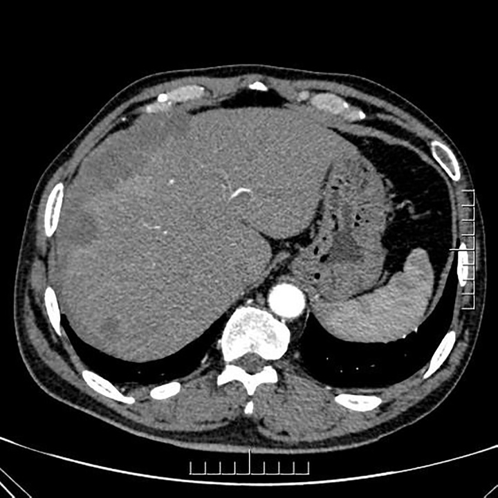
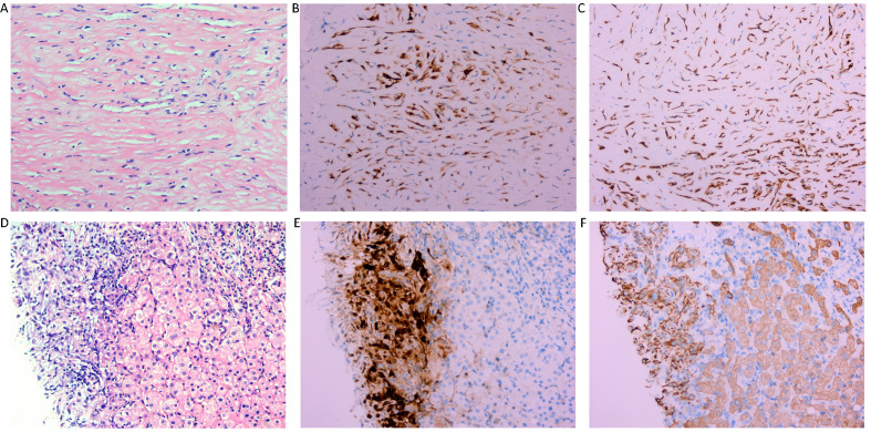
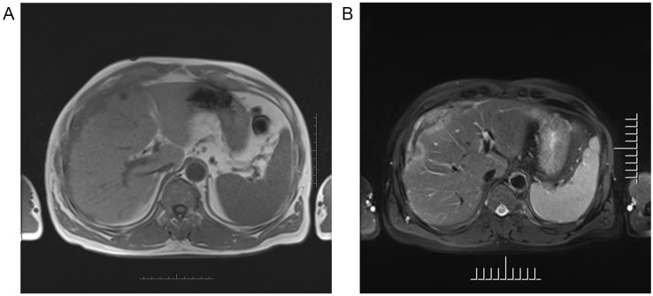
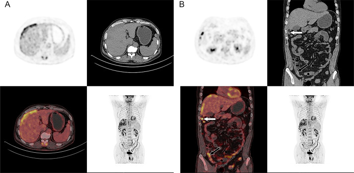
Desmoplastic malignant peritoneal mesothelioma (DMPM) is an extremely rare and aggressive subtype of sarcomatoid malignant mesothelioma, originating from the mesothelial lining of body cavities. It is characterized by significant local invasiveness and poor prognosis. The nonspecific symptoms of DMPM often result in delayed diagnosis. This case report presents the multimodality imaging findings of DMPM in a 58-year-old male, including ultrasound, CT, contrast-enhanced CT, magnetic resonance imaging (MRI), and 18-fluorodeoxy-glucose positron emission tomography combined with CT (18F-FDG PET/CT). These findings aim to enhance radiologists' understanding of the imaging features and differential diagnosis of DMPM. In this case, the tumor was located in the right subdiaphragm and the right anterior and left medial lobes of the liver. Due to the patient's history of alcoholic cirrhosis-a known risk factor for primary liver tumors-the initial diagnostic focus was on identifying a primary liver tumor with potential peritoneal invasion, overlooking other possible etiologies. However, histological results revealed that the liver lesion was secondary to invasion by DMPM. To the best of our knowledge, cases of DMPM invading the liver are exceedingly rare. This report underscores the importance of considering peritoneal tumors in the differential diagnosis when lesions involve both the peritoneum and adjacent organs, despite their rarity.

摘要

促纤维增生性恶性腹膜间皮瘤(DMPM)是肉瘤样恶性间皮瘤中一种极其罕见且侵袭性强的亚型,起源于体腔的间皮内衬。其特点是具有显著的局部侵袭性且预后较差。DMPM的非特异性症状常导致诊断延迟。本病例报告展示了一名58岁男性DMPM的多模态影像学表现,包括超声、CT、增强CT、磁共振成像(MRI)以及18-氟脱氧葡萄糖正电子发射断层扫描联合CT(18F-FDG PET/CT)。这些发现旨在提高放射科医生对DMPM影像学特征及鉴别诊断的认识。在该病例中,肿瘤位于右膈下以及肝脏的右前叶和左内侧叶。由于患者有酒精性肝硬化病史——原发性肝癌的已知危险因素——最初的诊断重点是识别可能伴有腹膜侵犯的原发性肝癌,而忽略了其他可能的病因。然而,组织学结果显示肝脏病变是由DMPM侵犯所致。据我们所知,DMPM侵犯肝脏的病例极为罕见。本报告强调,尽管腹膜肿瘤罕见,但当病变累及腹膜和邻近器官时,在鉴别诊断中考虑腹膜肿瘤的重要性。

https://cdn.ncbi.nlm.nih.gov/pmc/blobs/d509/11670191/407daa5744d8/fonc-14-1502105-g004.jpg
https://cdn.ncbi.nlm.nih.gov/pmc/blobs/d509/11670191/d85c035123b6/fonc-14-1502105-g001.jpg
https://cdn.ncbi.nlm.nih.gov/pmc/blobs/d509/11670191/41dd6a6cc1a8/fonc-14-1502105-g002.jpg
https://cdn.ncbi.nlm.nih.gov/pmc/blobs/d509/11670191/420da52cb5de/fonc-14-1502105-g003.jpg
https://cdn.ncbi.nlm.nih.gov/pmc/blobs/d509/11670191/407daa5744d8/fonc-14-1502105-g004.jpg

相似文献

1
Case report: Radiological features of a case of desmoplastic malignant mesothelioma of peritoneum.病例报告:一例腹膜促纤维增生性恶性间皮瘤的放射学特征
Front Oncol. 2024 Dec 11;14:1502105. doi: 10.3389/fonc.2024.1502105. eCollection 2024.
2
Enhanced Detection of Recurrent Diffuse Malignant Peritoneal Mesothelioma Using Ga-FAPI PET/CT Compared to F-FDG PET/CT: A Case Report.与¹⁸F-FDG PET/CT相比,¹⁸Ga-FAPI PET/CT在复发性弥漫性恶性腹膜间皮瘤检测中的应用:一例报告
World J Nucl Med. 2025 Mar 12;24(2):185-188. doi: 10.1055/s-0045-1805043. eCollection 2025 Jun.
3
CT differentiation of diffuse malignant peritoneal mesothelioma and peritoneal carcinomatosis.弥漫性恶性腹膜间皮瘤与腹膜转移癌的CT鉴别
J Gastroenterol Hepatol. 2016 Apr;31(4):709-15. doi: 10.1111/jgh.13260.
4
The value of BRCA-1-associated protein 1 expression and cyclin-dependent kinase inhibitor 2A deletion to distinguish peritoneal malignant mesothelioma from peritoneal location of carcinoma in effusion cytology specimens.BRCA-1 相关蛋白 1 表达和细胞周期蛋白依赖性激酶抑制剂 2A 缺失在鉴别腹腔渗出液细胞学标本中腹膜恶性间皮瘤与癌腹膜转移中的价值。
Cytopathology. 2020 Jan;31(1):5-11. doi: 10.1111/cyt.12788.
5
18F-FDG PET/CT in a recurrent diffuse malignant peritoneal mesothelioma.18F-FDG PET/CT 在复发性弥漫性恶性腹膜间皮瘤中的应用。
Clin Nucl Med. 2012 May;37(5):492-4. doi: 10.1097/RLU.0b013e3182478bb5.
6
Imaging of Malignant Pleural, Pericardial, and Peritoneal Mesothelioma.恶性胸膜、心包和腹膜间皮瘤的影像学表现。
Adv Anat Pathol. 2023 Jul 1;30(4):280-291. doi: 10.1097/PAP.0000000000000386. Epub 2022 Nov 18.
7
5-hmC loss is another useful tool in addition to BAP1 and MTAP immunostains to distinguish diffuse malignant peritoneal mesothelioma from reactive mesothelial hyperplasia in peritoneal cytology cell-blocks and biopsies.5-hmC 缺失除了 BAP1 和 MTAP 免疫染色之外,还是一种有用的工具,可用于在腹腔细胞学细胞块和活检中区分弥漫性恶性腹膜间皮瘤和反应性间皮增生。
Virchows Arch. 2022 Jul;481(1):23-29. doi: 10.1007/s00428-022-03336-1. Epub 2022 May 16.
8
Current concepts in the evaluation and treatment of patients with diffuse malignant peritoneal mesothelioma.弥漫性恶性腹膜间皮瘤患者评估和治疗的当前概念。
J Natl Compr Canc Netw. 2012 Jan;10(1):49-57. doi: 10.6004/jnccn.2012.0008.
9
Pleomorphic epithelioid diffuse malignant pleural mesothelioma: a clinicopathological review and conceptual proposal to reclassify as biphasic or sarcomatoid mesothelioma.多形性上皮样弥漫性恶性胸膜间皮瘤:临床病理回顾和重新分类为双相或肉瘤样间皮瘤的概念建议。
J Thorac Oncol. 2011 May;6(5):896-904. doi: 10.1097/JTO.0b013e318211127a.
10
Diffuse malignant peritoneal mesothelioma mimicking ovarian cancer.弥漫性恶性腹膜间皮瘤酷似卵巢癌。
Radiol Case Rep. 2021 Dec 28;17(3):779-783. doi: 10.1016/j.radcr.2021.11.021. eCollection 2022 Mar.

本文引用的文献

1
Diagnostic and Therapeutic Pathway in Diffuse Malignant Peritoneal Mesothelioma.弥漫性恶性腹膜间皮瘤的诊断与治疗路径
Cancers (Basel). 2023 Jan 21;15(3):662. doi: 10.3390/cancers15030662.
2
Peritoneal disease: key imaging findings that help in the differential diagnosis.腹膜疾病:有助于鉴别诊断的关键影像学表现。
Br J Radiol. 2022 Feb 1;95(1130):20210346. doi: 10.1259/bjr.20210346. Epub 2021 Nov 23.
3
Malignant mesothelioma: A histomorphological and immunohistochemical study of 24 cases from a tertiary care hospital in Southern India.
恶性间皮瘤:来自印度南部一家三级护理医院的24例病例的组织形态学和免疫组织化学研究
Indian J Cancer. 2018 Apr-Jun;55(2):190-195. doi: 10.4103/ijc.IJC_584_17.
4
Malignant peritoneal mesothelioma: a review.恶性腹膜间皮瘤:综述
Transl Lung Cancer Res. 2018 Oct;7(5):537-542. doi: 10.21037/tlcr.2018.10.04.
5
Clinical presentation, diagnosis, classification and management of peritoneal mesothelioma: a review.腹膜间皮瘤的临床表现、诊断、分类及管理:综述
J Gastrointest Oncol. 2017 Oct;8(5):915-924. doi: 10.21037/jgo.2017.08.01.
6
Guidelines for Pathologic Diagnosis of Malignant Mesothelioma 2017 Update of the Consensus Statement From the International Mesothelioma Interest Group.恶性间皮瘤病理诊断指南:国际间皮瘤兴趣小组共识声明2017年更新版
Arch Pathol Lab Med. 2018 Jan;142(1):89-108. doi: 10.5858/arpa.2017-0124-RA. Epub 2017 Jul 7.
7
18F-FDG-PET/CT of peritoneal tumors: a pictorial essay.18F-氟代脱氧葡萄糖正电子发射断层扫描/计算机断层扫描在腹膜肿瘤中的应用:图文综述
Nucl Med Commun. 2017 Jan;38(1):1-9. doi: 10.1097/MNM.0000000000000613.
8
CT differentiation of diffuse malignant peritoneal mesothelioma and peritoneal carcinomatosis.弥漫性恶性腹膜间皮瘤与腹膜转移癌的CT鉴别
J Gastroenterol Hepatol. 2016 Apr;31(4):709-15. doi: 10.1111/jgh.13260.
9
Introduction to The 2015 World Health Organization Classification of Tumors of the Lung, Pleura, Thymus, and Heart.《2015年世界卫生组织肺、胸膜、胸腺和心脏肿瘤分类》简介
J Thorac Oncol. 2015 Sep;10(9):1240-1242. doi: 10.1097/JTO.0000000000000663.
10
Desmoplastic malignant mesothelioma of the peritoneum.腹膜促纤维增生性恶性间皮瘤
Oxf Med Case Reports. 2014 Jun 14;2014(3):49-51. doi: 10.1093/omcr/omu021. eCollection 2014 Jun.