Wakabayashi Ryo, Azuma Seiichi, Hayashi Saori, Ueda Yuji, Iwakiri Masaki, Asamoto Masaaki, Uchida Kanji
Department of Anesthesiology and Pain Relief Center, The University of Tokyo Hospital, 7-3-1 Hongo, Bunkyo-ku, Tokyo, 113-8655, Japan.
JA Clin Rep. 2024 Dec 28;10(1):80. doi: 10.1186/s40981-024-00763-8.
Local anesthetic systemic toxicity (LAST) is a rare but potentially life-threatening complication. Under general anesthesia, neurological signs are often masked, delaying diagnosis and increasing the risk of sudden cardiovascular collapse. Therefore, early detection methods are critically needed.
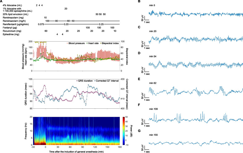
A 48-year-old male patient (height: 182 cm, weight: 98 kg) underwent resection of a mediastinal goiter. He received 10 mL of 4% lidocaine for topical airway anesthesia and 20 mL of 1% lidocaine with 1:100,000 epinephrine for chest wall anesthesia. Thirty minutes after airway anesthesia, continuous theta waves appeared on the frontal electroencephalogram (EEG), which were enhanced following chest wall anesthesia. These waves transitioned into a repeating pattern and evolved into sharp periodic discharges. After administering 150 mL of 20% lipid emulsion, the EEG normalized.
This case highlights that EEG monitoring during general anesthesia may facilitate the early detection of LAST and provide real-time feedback on treatment efficacy.
局部麻醉药全身毒性反应(LAST)是一种罕见但可能危及生命的并发症。在全身麻醉下,神经体征常被掩盖,延迟诊断并增加突然心血管崩溃的风险。因此,迫切需要早期检测方法。
一名48岁男性患者(身高:182厘米,体重:98千克)接受纵隔甲状腺肿切除术。他接受了10毫升4%利多卡因用于气道局部麻醉,以及20毫升1%利多卡因加1:100,000肾上腺素用于胸壁麻醉。气道麻醉30分钟后,额叶脑电图(EEG)上出现连续的θ波,胸壁麻醉后增强。这些波转变为重复模式并演变为尖锐的周期性放电。给予150毫升20%脂质乳剂后,脑电图恢复正常。
本病例表明,全身麻醉期间的脑电图监测可能有助于早期检测LAST,并提供关于治疗效果的实时反馈。