• 文献检索
  • 文档翻译
  • 深度研究
  • 学术资讯
  • Suppr Zotero 插件Zotero 插件
  • 邀请有礼
  • 套餐&价格
  • 历史记录
应用&插件
Suppr Zotero 插件Zotero 插件浏览器插件Mac 客户端Windows 客户端微信小程序
定价
高级版会员购买积分包购买API积分包
服务
文献检索文档翻译深度研究API 文档MCP 服务
关于我们
关于 Suppr公司介绍联系我们用户协议隐私条款
关注我们

Suppr 超能文献

核心技术专利:CN118964589B侵权必究
粤ICP备2023148730 号-1Suppr @ 2026

文献检索

告别复杂PubMed语法,用中文像聊天一样搜索,搜遍4000万医学文献。AI智能推荐,让科研检索更轻松。

立即免费搜索

文件翻译

保留排版,准确专业,支持PDF/Word/PPT等文件格式,支持 12+语言互译。

免费翻译文档

深度研究

AI帮你快速写综述,25分钟生成高质量综述,智能提取关键信息,辅助科研写作。

立即免费体验

脂肪细胞对甲状旁腺腺瘤超声评估的影响。

Impact of adipocytes on ultrasound evaluation of parathyroid adenomas.

作者信息

Fujimoto Tomoko, Hirokawa Mitsuyoshi, Suzuki Ayana, Oshita Maki, Yamaoka Hiroyuki, Fujishima Makoto, Onoda Naoyoshi, Miyauchi Akira, Akamizu Takashi

机构信息

Department of Clinical Laboratory, Kuma Hospital, 8-2-35 Shimoyamate-Dori, Chuo-Ku, Kobe, Hyogo, 650-0011, Japan.

Department of Diagnostic Pathology and Cytology, Kuma Hospital, Kobe, Hyogo, 650-0011, Japan.

出版信息

J Med Ultrason (2001). 2025 Apr;52(2):237-243. doi: 10.1007/s10396-024-01511-2. Epub 2024 Dec 28.

DOI:10.1007/s10396-024-01511-2
PMID:39731664
原文链接:https://pmc.ncbi.nlm.nih.gov/articles/PMC12018633/
Abstract

PURPOSE

Parathyroid lipoadenomas are difficult to recognize preoperatively; hence, they may remain undetected. Difficulty in recognition is thought to be due to the adipocytes present in the tumor. This study aimed to clarify the impact of adipocytes as a component of parathyroid adenomas on ultrasound evaluation.

METHODS

Eighteen parathyroid adenoma cases, in which the adipose tissue accounted for more than 10% of the tumors, were included in this study. Of these, five were consistent with lipoadenomas. Twenty-five consecutive patients with parathyroid adenoma without adipocytes were used as controls.

RESULTS

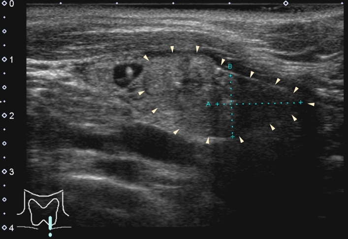
Ultrasonography revealed a lipoadenoma detection rate of 20.0%. This increased to 80.0% at re-examinations performed after obtaining information from other imaging modalities. Compared with parathyroid adenoma cases with no adipocytes or few adipocytes, the frequencies of ill-defined margins, iso- and/or hyperechogenicity, heterogeneous consistency with a two-tone pattern, poor vascular flow, no polar artery, and no hyperechoic line were significantly higher in parathyroid lipoadenoma cases. The hyperechoic and isoechoic areas in tumors with a two-tone pattern correspond to adipocyte- and parathyroid cell-rich areas, respectively. The lipoadenoma tumor sizes measured using ultrasound tended to be smaller than the actual sizes.

CONCLUSIONS

The characteristic ultrasound findings of lipoadenomas were clearly different from those of parathyroid adenomas with or without adipocytes. We believe that our findings may contribute to an increased detection rate of lipoadenomas and allow us to consider them in the differential diagnosis.

摘要

目的

甲状旁腺脂肪瘤术前难以识别,因此可能未被发现。识别困难被认为是由于肿瘤中存在脂肪细胞。本研究旨在阐明脂肪细胞作为甲状旁腺腺瘤的一个组成部分对超声评估的影响。

方法

本研究纳入了18例甲状旁腺腺瘤病例,其中脂肪组织占肿瘤的10%以上。其中5例符合脂肪瘤诊断。连续选取25例无脂肪细胞的甲状旁腺腺瘤患者作为对照。

结果

超声检查显示脂肪瘤的检出率为20.0%。在从其他影像学检查获得信息后进行的复查中,这一比例增至80.0%。与无脂肪细胞或脂肪细胞较少的甲状旁腺腺瘤病例相比,甲状旁腺脂肪瘤病例中边界不清、等回声和/或高回声、呈双色调模式的不均匀质地、血流差、无极动脉以及无高回声线的频率明显更高。双色调模式肿瘤中的高回声区和等回声区分别对应富含脂肪细胞和甲状旁腺细胞的区域。超声测量的脂肪瘤大小往往小于实际大小。

结论

脂肪瘤的特征性超声表现与有无脂肪细胞的甲状旁腺腺瘤明显不同。我们认为我们的研究结果可能有助于提高脂肪瘤的检出率,并使我们能够在鉴别诊断中考虑到它们。

https://cdn.ncbi.nlm.nih.gov/pmc/blobs/9136/12018633/a3c76e4cd518/10396_2024_1511_Fig5_HTML.jpg
https://cdn.ncbi.nlm.nih.gov/pmc/blobs/9136/12018633/5363d3019da0/10396_2024_1511_Fig1_HTML.jpg
https://cdn.ncbi.nlm.nih.gov/pmc/blobs/9136/12018633/9aa2af4428b4/10396_2024_1511_Fig2_HTML.jpg
https://cdn.ncbi.nlm.nih.gov/pmc/blobs/9136/12018633/344185ab3d4e/10396_2024_1511_Fig3_HTML.jpg
https://cdn.ncbi.nlm.nih.gov/pmc/blobs/9136/12018633/026deedb0942/10396_2024_1511_Fig4_HTML.jpg
https://cdn.ncbi.nlm.nih.gov/pmc/blobs/9136/12018633/a3c76e4cd518/10396_2024_1511_Fig5_HTML.jpg

相似文献

1
Impact of adipocytes on ultrasound evaluation of parathyroid adenomas.脂肪细胞对甲状旁腺腺瘤超声评估的影响。
J Med Ultrason (2001). 2025 Apr;52(2):237-243. doi: 10.1007/s10396-024-01511-2. Epub 2024 Dec 28.
2
A rare cause of primary hyperparathyroidism: Parathyroid lipoadenoma.原发性甲状旁腺功能亢进症的一种罕见病因:甲状旁腺脂肪瘤。
Auris Nasus Larynx. 2018 Dec;45(6):1245-1248. doi: 10.1016/j.anl.2018.05.001. Epub 2018 May 16.
3
Parathyroid lipoadenomas: a rare cause of primary hyperparathyroidism.甲状旁腺脂肪瘤:原发性甲状旁腺功能亢进的罕见病因。
Endocr Pract. 2006 Mar-Apr;12(2):131-6. doi: 10.4158/EP.12.2.131.
4
Pathology quiz case 2. Parathyroid lipoadenoma.
Arch Otolaryngol Head Neck Surg. 2006 Dec;132(12):1391-3. doi: 10.1001/archotol.132.12.1391.
5
VueBox® perfusion analysis of contrast-enhanced ultrasound (CEUS) examinations in patients with primary hyperparathyroidism for preoperative detection of parathyroid gland adenoma.VueBox®对原发性甲状旁腺功能亢进患者进行对比增强超声(CEUS)检查的灌注分析,用于术前检测甲状旁腺腺瘤。
Clin Hemorheol Microcirc. 2018;70(4):423-431. doi: 10.3233/CH-189307.
6
Two hundred consecutive parathyroid ultrasound studies by a single clinician: the impact of experience.
Endocr Pract. 2006 May-Jun;12(3):257-63. doi: 10.4158/EP.12.3.257.
7
Lipoadenoma of the parathyroid: characteristics of a rare cause of hyperparathyroidism.甲状旁腺脂肪腺瘤:甲状旁腺功能亢进的罕见病因特征。
Endocr J. 2022 Oct 28;69(10):1227-1232. doi: 10.1507/endocrj.EJ22-0118. Epub 2022 Jul 10.
8
Technetium 99m tetrofosmin parathyroid imaging. Results with double-phase study and SPECT in primary and secondary hyperparathyroidism.锝99m 四膦酸盐甲状旁腺显像。原发性和继发性甲状旁腺功能亢进的双期研究及单光子发射计算机断层扫描结果
Invest Radiol. 1997 Aug;32(8):459-65. doi: 10.1097/00004424-199708000-00005.
9
Evaluation of the microvascularization of pathologic parathyroid glands in patients with primary hyperparathyroidism using conventional ultrasound and contrast-enhanced ultrasound.应用常规超声和超声造影评估原发性甲状旁腺功能亢进症患者甲状旁腺病变的微血管化。
Clin Hemorheol Microcirc. 2011;48(1):95-103. doi: 10.3233/CH-2011-1402.
10
The role of colour Doppler ultrasonography in the preoperative localization of parathyroid adenomas.彩色多普勒超声在甲状旁腺瘤术前定位中的作用。
Endocr J. 2012;59(5):375-82. doi: 10.1507/endocrj.ej11-0351. Epub 2012 Feb 10.

本文引用的文献

1
Incidentally discovered parathyroid lipoadenoma in thyroid surgery: A case report and review of the literature.甲状腺手术中偶然发现的甲状旁腺脂肪瘤:一例报告并文献复习
Clin Case Rep. 2023 Nov 27;11(12):e8251. doi: 10.1002/ccr3.8251. eCollection 2023 Dec.
2
Lipoadenoma of the parathyroid: characteristics of a rare cause of hyperparathyroidism.甲状旁腺脂肪腺瘤:甲状旁腺功能亢进的罕见病因特征。
Endocr J. 2022 Oct 28;69(10):1227-1232. doi: 10.1507/endocrj.EJ22-0118. Epub 2022 Jul 10.
3
Overview of the 2022 WHO Classification of Parathyroid Tumors.
《2022 年世卫组织甲状旁腺肿瘤分类概述》。
Endocr Pathol. 2022 Mar;33(1):64-89. doi: 10.1007/s12022-022-09709-1. Epub 2022 Feb 17.
4
Atypical primary hyperparathyroidism due to parathyroid lipoadenoma: a case report.甲状旁腺脂肪瘤导致的非典型原发性甲状旁腺功能亢进:一例报告
J Surg Case Rep. 2021 Jul 23;2021(7):rjab308. doi: 10.1093/jscr/rjab308. eCollection 2021 Jul.
5
Parathyroid lipoadenoma: The particularities of a rare entity a case report and review of the literature.甲状旁腺脂肪瘤:一种罕见实体的特殊性——病例报告及文献复习
Int J Surg Case Rep. 2021 Feb;79:455-458. doi: 10.1016/j.ijscr.2021.01.091. Epub 2021 Jan 28.
6
Lipoadenoma of the Parathyroid Gland: Characterization of an Institutional Series Spanning 28 Years.甲状旁腺脂肪腺瘤:28 年的机构系列研究特征。
Endocr Pathol. 2020 Jun;31(2):156-165. doi: 10.1007/s12022-020-09616-3.
7
Ultrasound of Soft Tissue Masses and Fluid Collections.软组织肿块和液体积聚的超声检查
Radiol Clin North Am. 2019 May;57(3):657-669. doi: 10.1016/j.rcl.2019.01.013. Epub 2019 Feb 15.
8
Parathyroid Lipoadenoma: A Pitfall in Preoperative Localization.甲状旁腺脂肪瘤:术前定位中的一个陷阱。
Intern Med. 2019 Apr 15;58(8):1183-1184. doi: 10.2169/internalmedicine.1249-18. Epub 2018 Dec 18.
9
Phantom Nodules Detected by Ultrasound Examination of the Neck: The Possibility of Ectopic Cervical Thymic Tissue in Adults.颈部超声检查发现的假结节:成人异位颈胸腺组织的可能性
Ultrasound Int Open. 2018 Oct;4(4):E119-E123. doi: 10.1055/a-0747-6453. Epub 2018 Oct 19.
10
Doppler ultrasound appearance of neck tumors.颈部肿瘤的多普勒超声表现。
J Ultrason. 2018;18(73):96-102. doi: 10.15557/JoU.2018.0014.