Zhang Yan-Ting, Wang Sheng-Hui, Zhao Li, Wang Hui-Min, Wang Lin, Shi Rong-Rong, Liang Si-Chao, Li Bao-Feng, Chen Bei
Department of Radiation Oncology, Nanfang Hospital, Southern Medical University, 1838 North Guangzhou Avenue, Guangzhou, 510515, Guangdong, P.R. China.
Department of Cardiovascular Surgery, Nanfang Hospital, Southern Medical University, Guangzhou, 510515, China.
BMC Cancer. 2024 Dec 30;24(1):1595. doi: 10.1186/s12885-024-13375-3.
Antiangiogenesis therapy has become a hot field in cancer research. Given that tumor blood vessels often express specific markers related to angiogenesis, the study of these heterogeneous molecules in different tumor vessels holds promise for advancing anti-angiogenic therapy. Previously using phage display technology, we identified a targeting peptide named GX1 homing to gastric cancer vessels for the first time. However, GX1 also showed some non-specific binding with normal gastric vessels, which can lead to toxic side effects on normal endothelial cells. Therefore, we urgently need to adopt new screening strategies to avoid non-specific binding to normal vessels and obtain gastric cancer vascular targeting peptides with higher specificity.
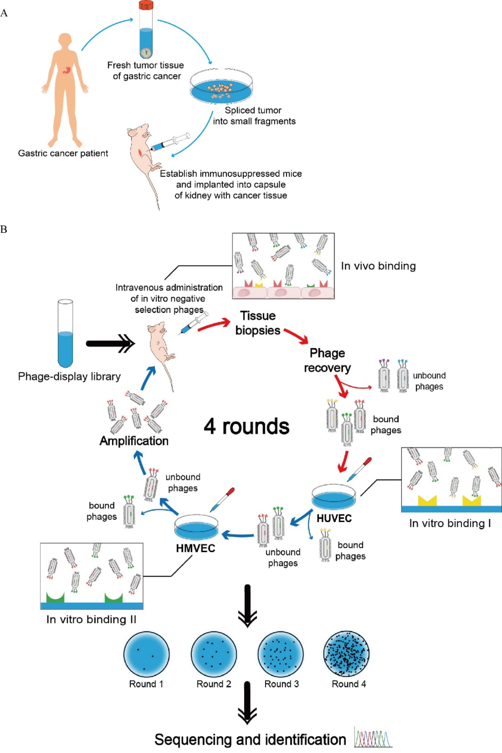
In this study, we designed a new strategy which combined "positive screening" in vivo and "negative screening" in vitro for the first time. An in vivo positive screening was conducted using tumor bearing nude mice to identify peptides that were specifically enriched within the vasculature of gastric cancer. Concurrently, an in vitro negative screening process was conducted on normal vasculature endothelial cells, including human umbilical vein endothelial cells (HUVECs) and human microvascular endothelial cells (HMVECs), to eliminate peptides binding to normal vasculature. After four rounds of iterative screening, a targeting peptide specifically targeting gastric cancer vasculature was obtained. In addition, an in vitro co-culture model by culturing HUVEC in tumor conditioned medium (Co-HUVEC) was established to investigate the affinity of these peptides. The targeting peptide was labeled with fluorescein isothiocyanate (FITC) for competitive and inhibitory assays.
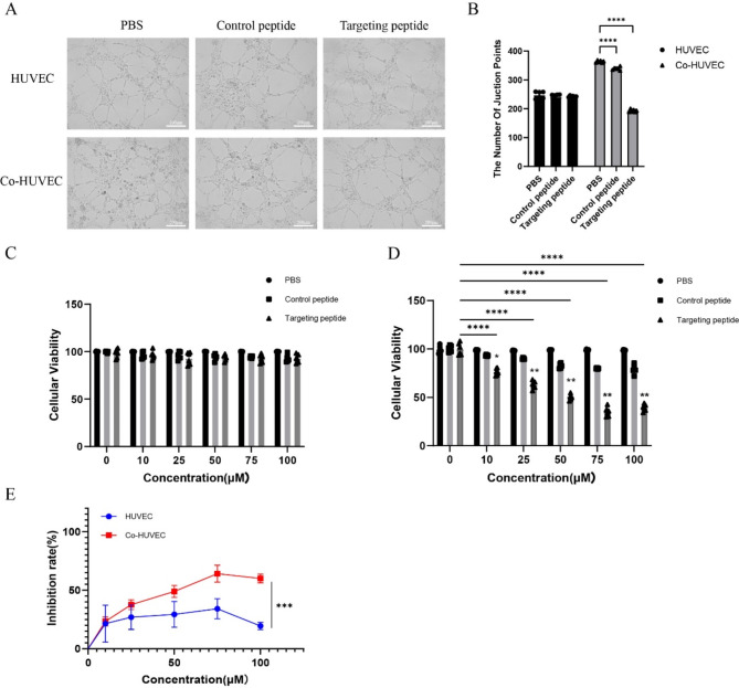
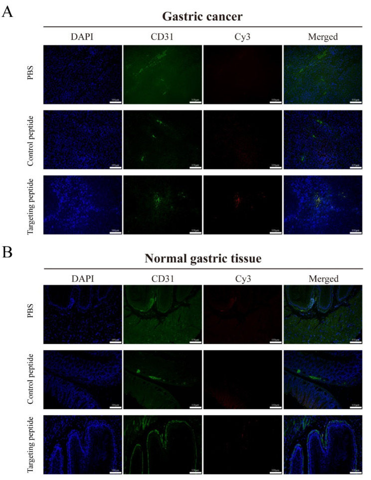
Blood vessel density analysis confirmed redundant capillary vessels in the xenografts, indicating that the mouse model was suitable for positive screening. Following four rounds of panning, a significant enrichment for phages specifically binding to gastric cancer vasculature was observed, with minimal binding to normal endothelial cells. The peptide CNTGSPYEC exhibited the highest reproducibility. In vitro immunofluorescence staining confirmed that the peptide CNTGSPYEC could specifically enrich in Co-HUVECs while showing no binding to normal vascular endothelial cells. In vivo immunofluorescence staining revealed that the peptide CNTGSPYEC could only bind to vascular endothelial cells specifically in gastric cancer but show no non-specific binding with normal tissue. Competitive and inhibitory assay also verified the targeting characteristics of the peptide with the fluorescence intensity of 17.13. As the concentration increases, the competitive inhibition rate can be incrementally raised to 93% (p < 0.05). Endothelial tube formation assay indicated that the peptide could suppress neovascularization, with the microvessel count reducing by 40% (p < 0.05). Furthermore, Cell Counting Kit-8 assay (CCK8) showed that the targeting peptide could partly inhibit cell proliferation of Co-HUVEC (61.7%).
Our novel strategy of the combined in vitro and in vivo screening outperforms previous methods that relied solely on negative/positive screening. In vivo and in vitro test confirmed the high targeting characteristic of the new peptide. Therefore, the peptide CNTGSPYEC may be a potential candidate in diagnosis and anti-angiogenesis therapy of gastric cancer. Our further exploration employs it as a vehicle for mediating drug accumulation in gastric cancer tissue.
抗血管生成疗法已成为癌症研究的一个热门领域。鉴于肿瘤血管通常表达与血管生成相关的特定标志物,研究不同肿瘤血管中的这些异质分子有望推动抗血管生成疗法的发展。此前我们利用噬菌体展示技术首次鉴定出一种靶向肽GX1,它可归巢至胃癌血管。然而,GX1也与正常胃血管表现出一些非特异性结合,这可能导致对正常内皮细胞产生毒副作用。因此,我们迫切需要采用新的筛选策略,以避免与正常血管的非特异性结合,并获得具有更高特异性的胃癌血管靶向肽。
在本研究中,我们首次设计了一种将体内“阳性筛选”和体外“阴性筛选”相结合的新策略。使用荷瘤裸鼠进行体内阳性筛选,以鉴定在胃癌脉管系统中特异性富集的肽。同时,对包括人脐静脉内皮细胞(HUVECs)和人微血管内皮细胞(HMVECs)在内的正常脉管系统内皮细胞进行体外阴性筛选过程,以消除与正常脉管系统结合的肽。经过四轮迭代筛选,获得了一种特异性靶向胃癌脉管系统的靶向肽。此外,通过在肿瘤条件培养基中培养HUVEC建立了体外共培养模型(Co-HUVEC),以研究这些肽的亲和力。将靶向肽用异硫氰酸荧光素(FITC)标记进行竞争和抑制试验。
血管密度分析证实异种移植瘤中存在多余的毛细血管,表明该小鼠模型适合进行阳性筛选。经过四轮淘选,观察到与胃癌脉管系统特异性结合的噬菌体显著富集,与正常内皮细胞的结合最少。肽CNTGSPYEC表现出最高的重复性。体外免疫荧光染色证实肽CNTGSPYEC可特异性富集于Co-HUVECs中,而与正常血管内皮细胞无结合。体内免疫荧光染色显示肽CNTGSPYEC仅能特异性结合胃癌中的血管内皮细胞,与正常组织无非特异性结合。竞争和抑制试验也验证了该肽的靶向特性,荧光强度为17.13。随着浓度增加,竞争抑制率可逐步提高至93%(p < 0.05)。内皮管形成试验表明该肽可抑制新血管形成,微血管数量减少40%(p < 0.05)。此外,细胞计数试剂盒-8检测(CCK8)显示靶向肽可部分抑制Co-HUVEC的细胞增殖(61.7%)。
我们新颖的体内外联合筛选策略优于以往仅依赖阴性/阳性筛选的方法。体内和体外试验证实了新肽的高靶向特性。因此,肽CNTGSPYEC可能是胃癌诊断和抗血管生成治疗的潜在候选物。我们进一步探索将其用作介导药物在胃癌组织中蓄积的载体。