Leonhardi Jakob, Schnarkowski Benedikt, Mehdorn Matthias, Höhn Anne-Kathrin, Niebisch Stefan, Plum Patrick, Seehofer Daniel, Tiepolt Solveig, Denecke Timm, Meyer Hans-Jonas
Department of Diagnostic and Interventional Radiology, University of Leipzig, Leipzig, Germany.
Department of Visceral and Transplantation Surgery, University Hospital Leipzig, University of Leipzig, Leipzig, Germany.
In Vivo. 2025 Jan-Feb;39(1):353-359. doi: 10.21873/invivo.13835.
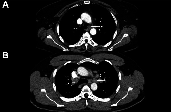
BACKGROUND/AIM: The recently published Node-Reporting and Data System (Node-RADS) can aid the characterization of lymph nodes in cross-sectional imaging. This study investigated the Node-RADS system in computed tomography (CT) to characterize lymph nodes in esophageal cancer.
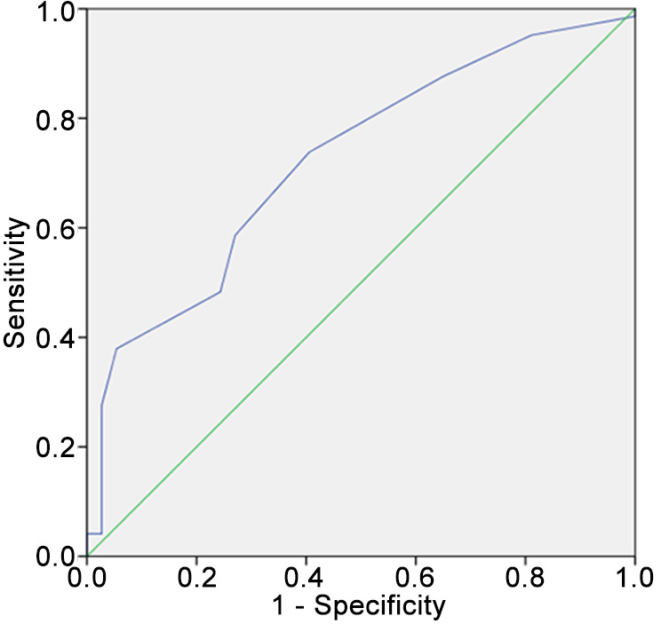
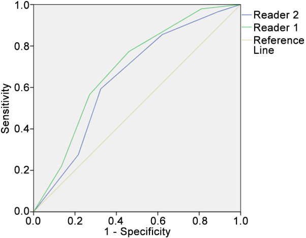
Overall, 126 patients (15 female, 11.9%) with a mean age of 62.1±10.4 years comprised the patient sample. All patients underwent resection with curative intent and the lymph nodes were histopathologically analyzed during clinical routine. For every patient, the locoregional lymph nodes were scored in accordance with the Node-RADS classification. For statistical analysis, receiver-operating characteristics (ROC) with area under the curve (AUC) were used to test for diagnostic accuracy; inter-reader variability was assessed with Cohen's kappa.
Overall, 54 patients were nodal positive (42.9%), 72 patients were nodal negative (57.1%). Inter-reader agreement was substantial for the overall Node-RADS scoring ([Formula: see text]=0.65, p<0.001). ROC curve analysis for lymph node discrimination (N0 versus N1-3) showed an AUC of 0.69 (95% confidence interval=0.59-0.79). A threshold score of more than 2 resulted in a sensitivity of 0.77 and a specificity of 0.55 for correctly predicting nodal positivity. Node-RADS 1 category had a malignancy rate of 30%, Node-RADS 2 of 14%, Node-RADS 3 of 81%, Node-RADS 4 of 90.1% and Node-RADS 5 of 86.5%.
The Node-RADS score on staging CT is associated with the malignancy rate of lymph nodes in patients with EC with only moderate diagnostic accuracy. The inter-reader variability is moderate, which could pose difficulties for translation into clinical routine.
背景/目的:最近发布的淋巴结报告与数据系统(Node-RADS)有助于在横断面成像中对淋巴结进行特征描述。本研究在计算机断层扫描(CT)中对Node-RADS系统进行研究,以对食管癌中的淋巴结进行特征描述。
总共126例患者(15例女性,占11.9%)纳入患者样本,平均年龄为62.1±10.4岁。所有患者均接受了根治性切除手术,并且在临床常规过程中对淋巴结进行了组织病理学分析。对于每位患者,根据Node-RADS分类对局部区域淋巴结进行评分。为进行统计分析,采用曲线下面积(AUC)的受试者操作特征(ROC)曲线来检验诊断准确性;采用Cohen's kappa评估阅片者间的变异性。
总体而言,54例患者淋巴结阳性(42.9%),72例患者淋巴结阴性(57.1%)。阅片者间对于总体Node-RADS评分的一致性较高([公式:见原文]=0.65,p<0.001)。淋巴结鉴别(N0与N1-3)的ROC曲线分析显示AUC为0.69(95%置信区间=0.59-0.79)。阈值分数大于2时,正确预测淋巴结阳性的敏感性为0.77,特异性为0.55。Node-RADS 1类的恶性率为30%,Node-RADS 2类为14%,Node-RADS 3类为81%,Node-RADS 4类为90.1%,Node-RADS 5类为86.5%。
分期CT上的Node-RADS评分与食管癌患者淋巴结的恶性率相关,但诊断准确性仅为中等。阅片者间的变异性为中等,这可能给转化为临床常规应用带来困难。