Wälchli Thomas, Bhatia Kartik Dev, Guest Will, Bisschop Jeroen, Olijnyk Leonardo, Kortman Hans, Constanthin Paul E, Nicholson Patrick, Monnier Philippe P, Kalyvas Aristotelis, Winkler Ethan A, Berhouma Moncef, Krings Timo, Radovanovic Ivan
Group Brain Vasculature and Perivascular Niche, Division of Experimental and Translational Neuroscience, Krembil Brain Institute, Krembil Research Institute, Toronto Western Hospital, University Health Network, University of Toronto, Toronto, ON, Canada;
Division of Neurosurgery, Department of Surgery, Toronto Western Hospital, University Health Network, University of Toronto, Toronto, ON, Canada.
In Vivo. 2025 Jan-Feb;39(1):280-291. doi: 10.21873/invivo.13826.
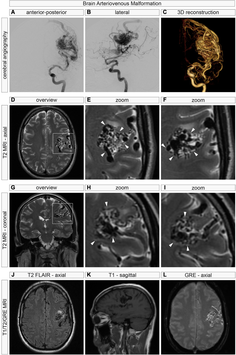
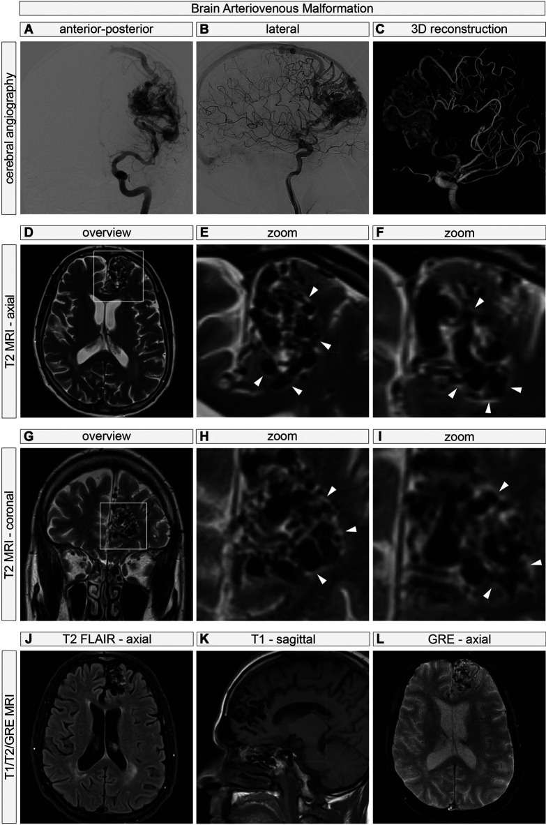
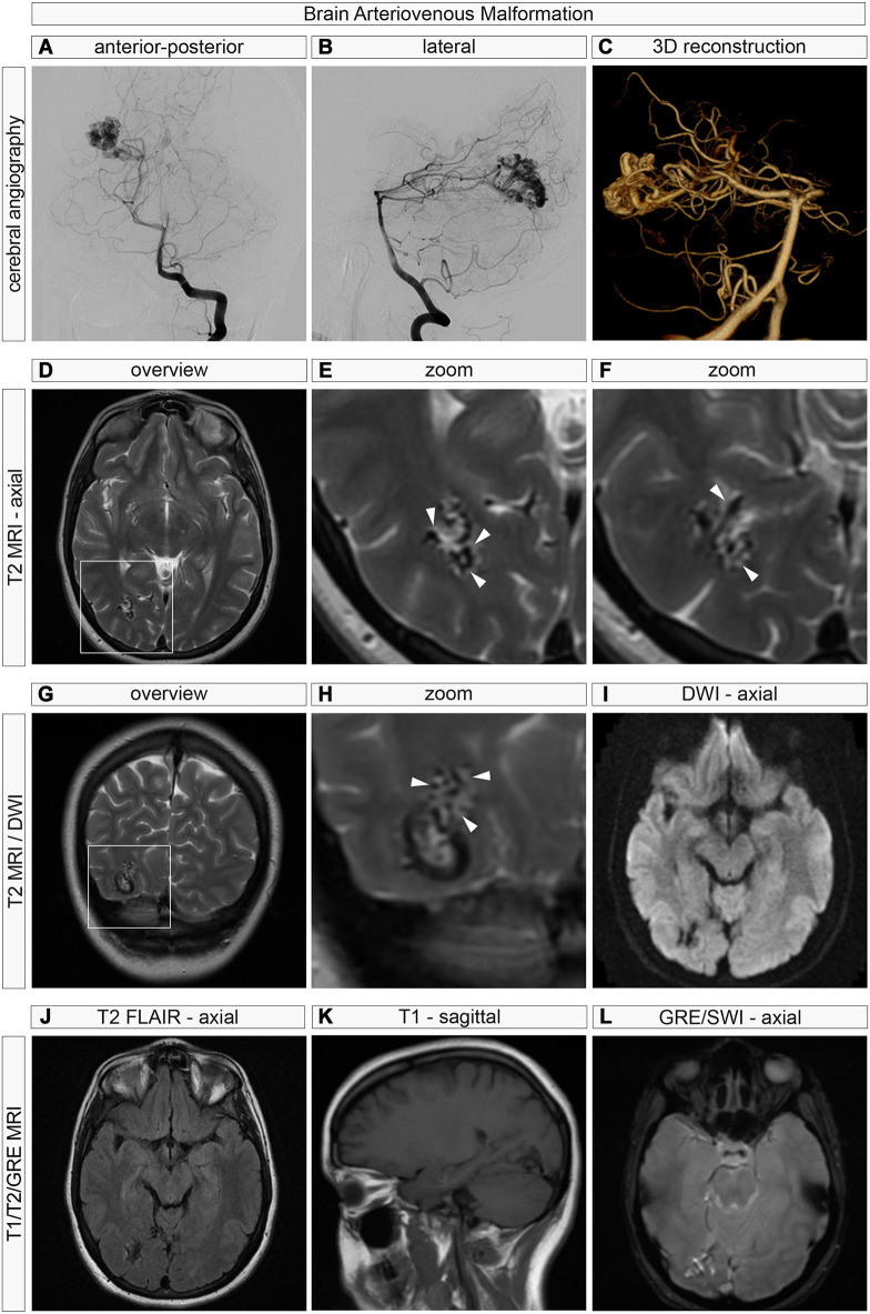
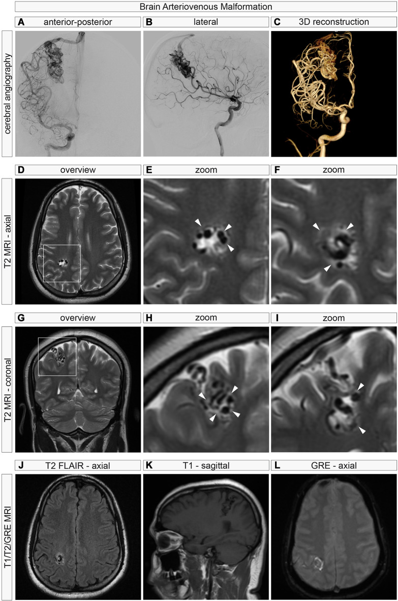
BACKGROUND/AIM: Brain arteriovenous malformations (AVMs) are vascular malformations characterized by dysmorphic, aberrant vasculature. During previous surgeries of compact nidus brain AVMs (representing the majority of cases), we have observed a "shiny" plane between nidal and perinidal AVM vessels and the surrounding grey and white matter and hypothesized that preoperative neuroimaging of brain AVMs may show a neuroradiological correlate of these intraoperative observations.
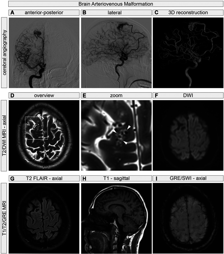
We retrospectively reviewed and analyzed multiplanar and multisequence 3-Tesla magnetic resonance (3T MR) imaging in five consecutive brain AVMs with special attention on imaging characteristics of the brain-AVM interface, i.e., the perivascular and perinidal regions.
In all five patients, we identified T2-hypertinense perivascular perinidal spaces, which were predominantly observed around the AVM nidus and less prominently around the feeding arteries or draining veins.
The identification of T2-hypertinense perivascular spaces surrounding brain AVMs on neuroradiological imaging may provide insights into the anatomico-radiological relationships of brain AVMs and the surrounding grey and white matter parenchyma. These findings could have future implications for our understanding of brain AVM biology and may influence neurosurgical approaches to these lesions.
背景/目的:脑动静脉畸形(AVM)是一种血管畸形,其特征为血管形态异常、走行紊乱。在以往对致密巢状脑AVM(占大多数病例)的手术中,我们观察到巢状和巢周AVM血管与周围灰质和白质之间存在一个“发亮”平面,并推测脑AVM的术前神经影像学检查可能显示出与这些术中观察结果相关的神经放射学表现。
我们回顾性分析了连续5例脑AVM的多平面、多序列3特斯拉磁共振(3T MR)成像,特别关注脑-AVM界面即血管周围和巢周区域的成像特征。
在所有5例患者中,我们均发现了T2高信号的血管周围巢周间隙,主要见于AVM巢周围,在供血动脉或引流静脉周围则不太明显。
在神经放射学成像中识别出脑AVM周围T2高信号的血管周围间隙,可能有助于深入了解脑AVM与周围灰质和白质实质之间的解剖学-放射学关系。这些发现可能对我们理解脑AVM生物学具有未来意义,并可能影响对这些病变的神经外科手术方法。