Tsuge Itaru, Saito Susumu, Munisso Maria Chiara, Kosaka Tomoko, Takaya Ayako, Liu Chang, Yamamoto Goshiro, Morimoto Naoki
Department of Plastic and Reconstructive Surgery, Graduate School of Medicine, Kyoto University, Kyoto, Japan.
Department of Medical Informatics, Graduate School of Medicine, Kyoto University, Kyoto, Japan.
Microsurgery. 2025 Jan;45(1):e70013. doi: 10.1002/micr.70013.
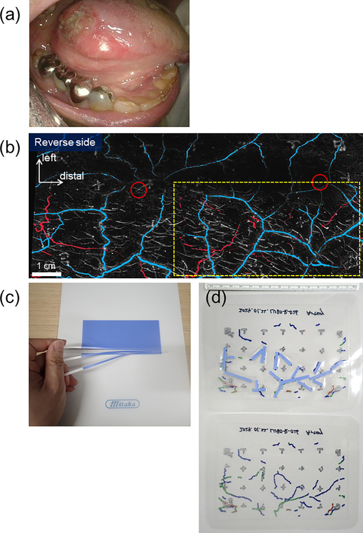
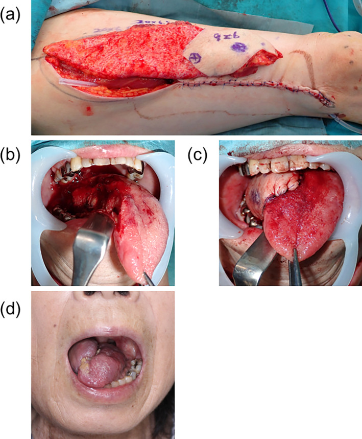
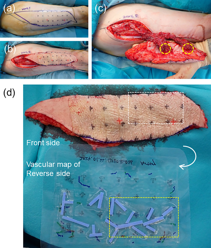
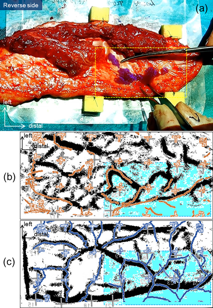
Thinning of anterolateral thigh flap is challenging. Anatomical studies have shown variations in arterial branching patterns in the subcutaneous layer, which were suspected to be the reason for the high frequency of thinning failures. We attempted to visualize subcutaneous arterial courses preoperatively and perform thinning of perforator flaps using this information appropriately. We accumulated evidence on the accuracy of noninvasive vascular visualization using photoacoustic tomography (PAT). In the present case, we applied a medical imaging projection system (MIPS), which enabled real-time surgical navigation using indocyanine green (ICG) emission signals, to use photoacoustic information intraoperatively during the flap thinning procedure. A 69-year-old woman underwent half-tongue resection using the pull-through method for right-sided tongue cancer. Preoperative PAT was performed 5 days before surgery. The 12 × 6-cm area took ~8 min to scan. We used an ICG test card containing ICG-positive control material cut into strips to show tentative artery lines by projection mapping. The transparent vascular map was laminated and sterilized. MIPS captured ICG fluorescence signals that penetrated the anterolateral thigh flap and continuously projected the purple area on the reverse side of the flap, guiding the position of the tentative arteries. A 20 × 6.5-cm anterolateral thigh flap was elevated with the distal part of the reconstructed tongue and proximal de-epithelialized part to fill the pull-thorough tunnel in the submandibular region. Active bleeding was observed when cutting marginal fat tissue near the purple line of the distal ALT flap projected by MIPS. The study protocol did not include a highly invasive trial for MIPS-guided thinning; therefore, we removed minimal marginal fat tissue. The ALT flap showed no postoperative complications while maintaining conversation and swallowing functions. We present the concept of subcutaneous arterial real-time navigation surgery using PAT and MIPS for safe, easy, and fast flap thinning procedures in the future.
大腿前外侧皮瓣的减薄具有挑战性。解剖学研究表明,皮下层动脉分支模式存在变异,这被怀疑是减薄失败频率高的原因。我们试图在术前可视化皮下动脉走行,并利用这些信息适当地进行穿支皮瓣的减薄。我们积累了关于使用光声断层扫描(PAT)进行无创血管可视化准确性的证据。在本病例中,我们应用了一种医学成像投影系统(MIPS),该系统能够利用吲哚菁绿(ICG)发射信号进行实时手术导航,以便在皮瓣减薄过程中术中使用光声信息。一名69岁女性因右侧舌癌采用牵拉法进行半舌切除术。术前5天进行了PAT检查。12×6厘米的区域扫描耗时约8分钟。我们使用了一张含有切成条状的ICG阳性对照材料的ICG测试卡,通过投影映射显示暂定动脉线。将透明血管图进行层压和消毒。MIPS捕捉穿透大腿前外侧皮瓣的ICG荧光信号,并在皮瓣背面持续投影紫色区域,引导暂定动脉的位置。将一个20×6.5厘米的大腿前外侧皮瓣与重建舌的远端部分和近端去上皮部分一起掀起,以填充下颌下区域的牵拉隧道。在切除MIPS投影的远端ALT皮瓣紫色线附近的边缘脂肪组织时观察到活动性出血。研究方案不包括MIPS引导下减薄的高侵入性试验;因此,我们仅切除了极少的边缘脂肪组织。ALT皮瓣术后未出现并发症,同时保持了言语和吞咽功能。我们提出了使用PAT和MIPS进行皮下动脉实时导航手术的概念,以便未来进行安全、简便和快速的皮瓣减薄手术。