Shamas Salman, Rahil Razia Rashid, Kaushal Laveena, Sharma Vinod Kumar, Wani Nissar Ahmad, Qureshi Shabir H, Ahmad Sheikh F, Attia Sabry M, Zargar Mohammad Afzal, Hamid Abid, Bhat Owais Mohmad
Department of Biotechnology, School of Life Sciences, Central University of Kashmir, Ganderbal 191201, India.
Department of Dermatology, Venereology & Leprology, Postgraduate Institute for Medical Education and Research, Chandigarh 160012, India.
Pharmaceuticals (Basel). 2024 Nov 22;17(12):1568. doi: 10.3390/ph17121568.
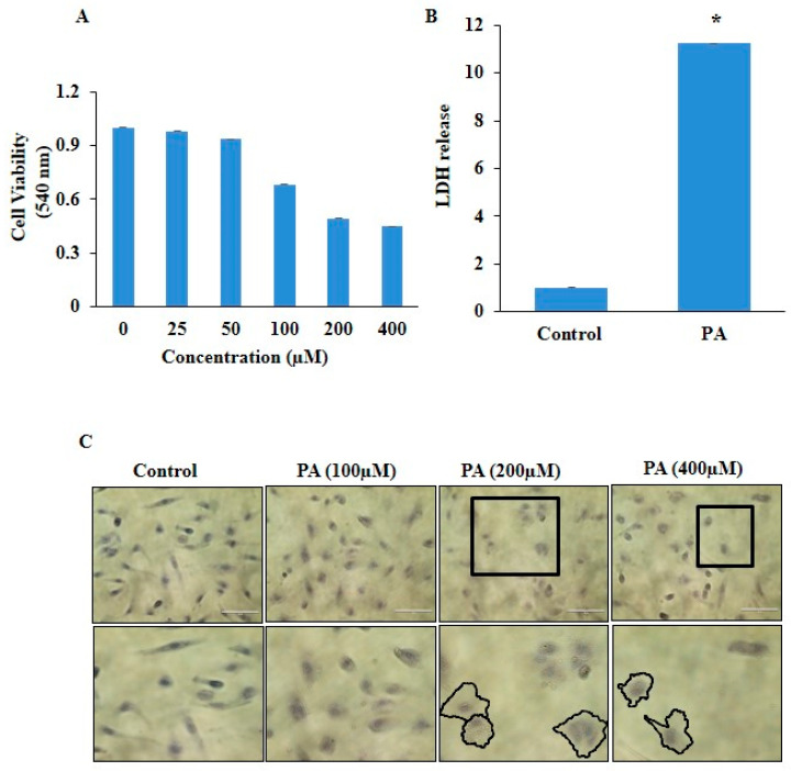
: Pyroptosis, an inflammatory cell death, is involved in the progression of atherosclerosis. Pyroptosis in endothelial cells (ECs) and its underlying mechanisms in atherosclerosis are poorly understood. Here, we investigated the role of a caspase-4/5-NF-κB pathway in pyroptosis in palmitic acid (PA)-stimulated ECs and EVs as players in pyroptosis. : Human umbilical vein endothelial cells (HUVECs) were cultured in an endothelial cell medium, treated with Ox-LDL, PA, caspase-4/5 inhibitor, NF-κB inhibitor, and sEV release inhibitor for 24 h, respectively. The cytotoxicity of PA was determined using an MTT assay, cell migration using a scratch-wound-healing assay, cell morphology using bright field microscopy, and lipid deposition using oil red O staining. The mRNA and protein expression of GSDM-D, CASP4, CASP5, NF-κB, NLRP3, IL-1β, and IL-18 were determined with RT-PCR and Western blot. Immunofluorescence was used to determine NLRP3 and ICAM-1 expressions. Extracellular vesicles (EVs) were isolated using an exosome isolation kit and were characterized by Western blot and scanning electron microscopy. PA stimulation significantly changed the morphology of the HUVECs characterized by cell swelling, plasma membrane rupture, and increased LDH release, which are features of pyroptosis. PA significantly increased lipid accumulation and reduced cell migration. PA also triggered inflammation and endothelial dysfunction, as evidenced by NLRP3 activation, upregulation of ICAM-1 (endothelial activation marker), and pyroptotic markers (NLRP3, GSDM-D, IL-1β, IL-18). Inhibition of caspase-4/5 (Ac-FLTD-CMK) and NF-κB (trifluoroacetate salt (TFA)) resulted in a significant reduction in LDH release and expression of caspase-4/5, NF-κB, and gasdermin D (GSDM-D) in PA-treated HUVECs. Furthermore, GW4869, an exosome release inhibitor, markedly reduced LDH release in PA-stimulated HUVECs. EVs derived from PA-treated HUVECs exacerbated pyroptosis, as indicated by significantly increased LDH release and augmented expression of GSDM-D, NF-κB. The present study revealed that inflammatory, non-canonical caspase-4/5-NF-κB signaling may be one of the crucial mechanistic pathways associated with pyroptosis in ECs, and pyroptotic EVs facilitated pyroptosis in normal ECs during atherosclerosis.
细胞焦亡是一种炎症性细胞死亡,参与动脉粥样硬化的进展。内皮细胞(ECs)中的细胞焦亡及其在动脉粥样硬化中的潜在机制尚不清楚。在此,我们研究了半胱天冬酶-4/5-核因子κB(NF-κB)信号通路在棕榈酸(PA)刺激的ECs和细胞外囊泡(EVs)焦亡中的作用,EVs是细胞焦亡的参与者。
人脐静脉内皮细胞(HUVECs)在内皮细胞培养基中培养,分别用氧化型低密度脂蛋白(Ox-LDL)、PA、半胱天冬酶-4/5抑制剂、NF-κB抑制剂和sEV释放抑制剂处理24小时。使用MTT法测定PA的细胞毒性,使用划痕愈合试验测定细胞迁移,使用明场显微镜观察细胞形态,使用油红O染色测定脂质沉积。用逆转录聚合酶链反应(RT-PCR)和蛋白质免疫印迹法测定GSDM-D、CASP4、CASP5、NF-κB、NLRP3、白细胞介素-1β(IL-1β)和白细胞介素-18(IL-18)的mRNA和蛋白质表达。用免疫荧光法测定NLRP3和细胞间黏附分子-1(ICAM-1)的表达。使用外泌体分离试剂盒分离细胞外囊泡,并通过蛋白质免疫印迹法和扫描电子显微镜进行鉴定。
PA刺激显著改变了HUVECs的形态,其特征为细胞肿胀、质膜破裂和乳酸脱氢酶(LDH)释放增加,这些都是细胞焦亡的特征。PA显著增加脂质积累并减少细胞迁移。PA还引发炎症和内皮功能障碍,NLRP3激活、ICAM-1(内皮激活标志物)上调以及细胞焦亡标志物(NLRP3、GSDM-D、IL-1β、IL-18)的表达均证明了这一点。抑制半胱天冬酶-4/5(Ac-FLTD-CMK)和NF-κB(三氟乙酸盐(TFA))可导致PA处理的HUVECs中LDH释放以及半胱天冬酶-4/5、NF-κB和gasdermin D(GSDM-D)的表达显著降低。此外,外泌体释放抑制剂GW4869显著降低了PA刺激的HUVECs中的LDH释放。PA处理的HUVECs来源的EVs加剧了细胞焦亡,表现为LDH释放显著增加以及GSDM-D、NF-κB的表达增强。
本研究表明,炎症性非经典半胱天冬酶-4/5-NF-κB信号通路可能是与ECs焦亡相关的关键机制途径之一,并且在动脉粥样硬化过程中,细胞焦亡性EVs促进了正常ECs的细胞焦亡。