Jeon Yunkyung, Kim Doohwa, Kim Mijin, Kim Bo Hyun, Pak Kyoungjune, Kim Jihyun, Kim Keunyoung
Division of Endocrinology, Department of Internal Medicine, Pusan National University Hospital, Busan, Republic of Korea.
Biomedical Research Institute, Pusan National University Hospital, Busan, Republic of Korea.
BMC Endocr Disord. 2025 Jan 8;25(1):6. doi: 10.1186/s12902-024-01800-4.
Despite TSH suppressive therapy improve the prognosis for the patient with differentiated thyroid cancer (DTC), there is an increasing concern regarding the potentially harmful effects of lifelong TSH suppression. Therefore, we aimed to examine the changes in body composition under TSH suppression in postmenopausal women with DTC.
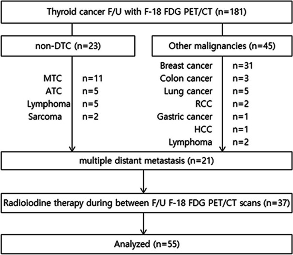
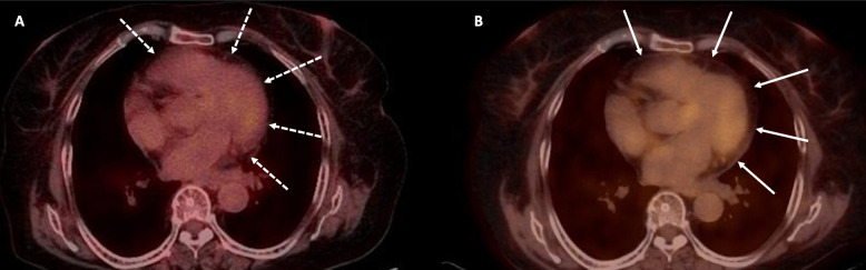
The body composition was assessed by the volumes as following; fat tissues of the epicardium and abdominal visceral and subcutaneous areas; bilateral psoas muscle or thigh muscle. Each volumetric measurements were performed using computed tomography (CT) scans using baseline and follow-up fluorine-18 fluorodeoxyglucose positron emission tomography/CT (F-FDG PET/CT)s for 2-year follow up period in Pusan National University Hospital, South Korea.
The 43 patients' median age was 50.0 years, and median body mass index (BMI) was 23.53 (interquartile range[IQR]: 22.19- 24.92) at the initial F-FDG PET/CT. The median follow-up period was 19.24 months (IQR: 17.24-21.79). No significant change in weight or BMI were observed during follow-up. Volumes of fat and muscles was not changed significantly except epicardial fat volume. The epicardial fat volume significantly increased during the follow-up period. The epicardial fat volumes were correlated with visceral fat volume, respectively, however, the changing ratio was only correlated with TSH suppression on multiple regression analysis.
Both skeletal muscle and abdominal fat volumes did not change, whereas epicardial fat volume increased over less than 2 years of observation under TSH suppressive therapy. Further research is needed for the harmonization of benefits or losses with the optimal TSH concentration in postmenopausal women.
尽管促甲状腺激素(TSH)抑制疗法可改善分化型甲状腺癌(DTC)患者的预后,但人们越来越关注终身TSH抑制的潜在有害影响。因此,我们旨在研究DTC绝经后女性在TSH抑制状态下身体成分的变化。
通过测量以下部位的体积来评估身体成分;心外膜、腹部内脏和皮下区域的脂肪组织;双侧腰大肌或大腿肌肉。在韩国釜山国立大学医院,使用基线和随访氟-18氟脱氧葡萄糖正电子发射断层扫描/计算机断层扫描(F-FDG PET/CT)进行为期2年的随访,通过计算机断层扫描(CT)扫描进行每次体积测量。
43例患者的中位年龄为50.0岁,初始F-FDG PET/CT时的中位体重指数(BMI)为23.53(四分位间距[IQR]:22.19-24.92)。中位随访期为19.24个月(IQR:17.24-21.79)。随访期间未观察到体重或BMI有显著变化。除心外膜脂肪体积外,脂肪和肌肉体积无显著变化。随访期间心外膜脂肪体积显著增加。心外膜脂肪体积分别与内脏脂肪体积相关,然而,在多元回归分析中,变化率仅与TSH抑制相关。
在TSH抑制治疗下观察不到2年的时间里,骨骼肌和腹部脂肪体积均未改变,而心外膜脂肪体积增加。需要进一步研究以平衡绝经后女性最佳TSH浓度的利弊。