• 文献检索
  • 文档翻译
  • 深度研究
  • 学术资讯
  • Suppr Zotero 插件Zotero 插件
  • 邀请有礼
  • 套餐&价格
  • 历史记录
应用&插件
Suppr Zotero 插件Zotero 插件浏览器插件Mac 客户端Windows 客户端微信小程序
定价
高级版会员购买积分包购买API积分包
服务
文献检索文档翻译深度研究API 文档MCP 服务
关于我们
关于 Suppr公司介绍联系我们用户协议隐私条款
关注我们

Suppr 超能文献

核心技术专利:CN118964589B侵权必究
粤ICP备2023148730 号-1Suppr @ 2026

文献检索

告别复杂PubMed语法,用中文像聊天一样搜索,搜遍4000万医学文献。AI智能推荐,让科研检索更轻松。

立即免费搜索

文件翻译

保留排版,准确专业,支持PDF/Word/PPT等文件格式,支持 12+语言互译。

免费翻译文档

深度研究

AI帮你快速写综述,25分钟生成高质量综述,智能提取关键信息,辅助科研写作。

立即免费体验

SEE:一种基于单细胞基因表达预测染色质构象动力学的方法。

SEE: A Method for Predicting the Dynamics of Chromatin Conformation Based on Single-Cell Gene Expression.

作者信息

Li Minghong, Yang Yurong, Wu Rucheng, Gong Haiyan, Yuan Zan, Wang Jixin, Long Erping, Zhang Xiaotong, Chen Yang

机构信息

State Key Laboratory of Common Mechanism Research for Major Diseases, Department of Biochemistry and Molecular Biology, Institute of Basic Medical Sciences, Chinese Academy of Medical Sciences and Peking Union Medical College, Beijing, 100005, China.

Department of Computer Science and Technology, University of Science and Technology Beijing, Beijing, 100083, China.

出版信息

Adv Sci (Weinh). 2025 Feb;12(8):e2406413. doi: 10.1002/advs.202406413. Epub 2025 Jan 7.

DOI:10.1002/advs.202406413
PMID:39778075
原文链接:https://pmc.ncbi.nlm.nih.gov/articles/PMC11848634/
Abstract

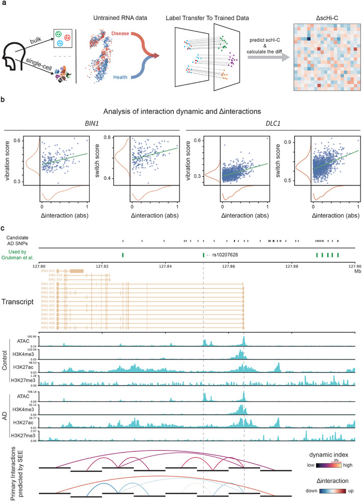
The dynamics of chromatin conformation involve continuous and reversible changes within the nucleus of a cell, which participate in regulating processes such as gene expression, DNA replication, and damage repair. Here, SEE is introduced, an artificial intelligence (AI) method that utilizes autoencoder and transformer techniques to analyze chromatin dynamics using single-cell RNA sequencing data and a limited number of single-cell Hi-C maps. SEE is employed to investigate chromatin dynamics across different scales, enabling the detection of (i) rearrangements in topologically associating domains (TADs), and (ii) oscillations in chromatin interactions at gene loci. Additionally, SEE facilitates the interpretation of disease-associated single-nucleotide polymorphisms (SNPs) by leveraging the dynamic features of chromatin conformation. Overall, SEE offers a single-cell, high-resolution approach to analyzing chromatin dynamics in both developmental and disease contexts.

摘要

染色质构象动力学涉及细胞内细胞核内连续且可逆的变化,这些变化参与调节基因表达、DNA复制和损伤修复等过程。本文介绍了SEE,这是一种人工智能(AI)方法,它利用自动编码器和变换器技术,通过单细胞RNA测序数据和有限数量的单细胞Hi-C图谱来分析染色质动力学。SEE用于研究不同尺度上的染色质动力学,能够检测(i)拓扑相关结构域(TADs)中的重排,以及(ii)基因位点处染色质相互作用的振荡。此外,SEE通过利用染色质构象的动态特征,有助于解释与疾病相关的单核苷酸多态性(SNPs)。总体而言,SEE提供了一种单细胞、高分辨率的方法,用于在发育和疾病背景下分析染色质动力学。

https://cdn.ncbi.nlm.nih.gov/pmc/blobs/eb2d/11848634/c6762d4ad1db/ADVS-12-2406413-g007.jpg
https://cdn.ncbi.nlm.nih.gov/pmc/blobs/eb2d/11848634/9f0cb2a966d2/ADVS-12-2406413-g002.jpg
https://cdn.ncbi.nlm.nih.gov/pmc/blobs/eb2d/11848634/f71e1b7b4fa5/ADVS-12-2406413-g003.jpg
https://cdn.ncbi.nlm.nih.gov/pmc/blobs/eb2d/11848634/f91f2d5ecd68/ADVS-12-2406413-g006.jpg
https://cdn.ncbi.nlm.nih.gov/pmc/blobs/eb2d/11848634/52d36219cb4a/ADVS-12-2406413-g008.jpg
https://cdn.ncbi.nlm.nih.gov/pmc/blobs/eb2d/11848634/95bb95f600e4/ADVS-12-2406413-g010.jpg
https://cdn.ncbi.nlm.nih.gov/pmc/blobs/eb2d/11848634/c6762d4ad1db/ADVS-12-2406413-g007.jpg

相似文献

1
SEE: A Method for Predicting the Dynamics of Chromatin Conformation Based on Single-Cell Gene Expression.SEE:一种基于单细胞基因表达预测染色质构象动力学的方法。
Adv Sci (Weinh). 2025 Feb;12(8):e2406413. doi: 10.1002/advs.202406413. Epub 2025 Jan 7.
2
Identifying topologically associating domains using differential kernels.使用差分核识别拓扑关联域。
PLoS Comput Biol. 2024 Jul 15;20(7):e1012221. doi: 10.1371/journal.pcbi.1012221. eCollection 2024 Jul.
3
The distributions of protein coding genes within chromatin domains in relation to human disease.染色质域内与人类疾病相关的蛋白质编码基因的分布。
Epigenetics Chromatin. 2019 Dec 5;12(1):72. doi: 10.1186/s13072-019-0317-2.
4
Single-nucleus Hi-C reveals unique chromatin reorganization at oocyte-to-zygote transition.单核Hi-C技术揭示了从卵母细胞到合子转变过程中独特的染色质重排。
Nature. 2017 Apr 6;544(7648):110-114. doi: 10.1038/nature21711. Epub 2017 Mar 29.
5
SnapHiC: a computational pipeline to identify chromatin loops from single-cell Hi-C data.SnapHiC:一种从单细胞 Hi-C 数据中识别染色质环的计算流程。
Nat Methods. 2021 Sep;18(9):1056-1059. doi: 10.1038/s41592-021-01231-2. Epub 2021 Aug 26.
6
Topologically associating domains are stable units of replication-timing regulation.拓扑相关结构域是复制时间调控的稳定单元。
Nature. 2014 Nov 20;515(7527):402-5. doi: 10.1038/nature13986.
7
Methods for the Analysis of Topologically Associating Domains (TADs).分析拓扑关联结构域(TADs)的方法。
Methods Mol Biol. 2022;2301:39-59. doi: 10.1007/978-1-0716-1390-0_3.
8
Microscopy-Based Chromosome Conformation Capture Enables Simultaneous Visualization of Genome Organization and Transcription in Intact Organisms.基于显微镜的染色体构象捕获技术使我们能够在完整的生物体中同时观察基因组组织和转录。
Mol Cell. 2019 Apr 4;74(1):212-222.e5. doi: 10.1016/j.molcel.2019.01.011. Epub 2019 Feb 19.
9
Evaluation of 3D Chromatin Interactions Using Hi-C.使用 Hi-C 技术评估 3D 染色质相互作用。
Methods Mol Biol. 2020;2117:65-78. doi: 10.1007/978-1-0716-0301-7_3.
10
Structural basis of differential gene expression at eQTLs loci from high-resolution ensemble models of 3D single-cell chromatin conformations.来自三维单细胞染色质构象高分辨率整合模型的eQTL位点差异基因表达的结构基础。
Bioinformatics. 2025 Feb 4;41(2). doi: 10.1093/bioinformatics/btaf050.

引用本文的文献

1
Topologically associating domains of chromatin on single-cell Hi-C data: a survey of bioinformatic tools and applications in the light of artificial intelligence.基于单细胞Hi-C数据的染色质拓扑相关结构域:人工智能视角下生物信息学工具及应用综述
Front Genet. 2025 Jul 1;16:1602234. doi: 10.3389/fgene.2025.1602234. eCollection 2025.

本文引用的文献

1
GAGE-seq concurrently profiles multiscale 3D genome organization and gene expression in single cells.GAGE-seq 可同时在单细胞中对多尺度 3D 基因组结构和基因表达进行分析。
Nat Genet. 2024 Aug;56(8):1701-1711. doi: 10.1038/s41588-024-01745-3. Epub 2024 May 14.
2
Simultaneous single-cell three-dimensional genome and gene expression profiling uncovers dynamic enhancer connectivity underlying olfactory receptor choice.同步单细胞三维基因组和基因表达谱分析揭示了嗅觉受体选择背后的动态增强子连接性。
Nat Methods. 2024 Jun;21(6):974-982. doi: 10.1038/s41592-024-02239-0. Epub 2024 Apr 15.
3
Fork coupling directs DNA replication elongation and termination.
叉形偶联指导DNA复制的延伸和终止。
Science. 2024 Mar 15;383(6688):1215-1222. doi: 10.1126/science.adj7606. Epub 2024 Mar 14.
4
Direct observation of a condensate effect on super-enhancer controlled gene bursting.直接观察凝聚物对超级增强子控制的基因爆发的影响。
Cell. 2024 Jan 18;187(2):331-344.e17. doi: 10.1016/j.cell.2023.12.005. Epub 2024 Jan 8.
5
Simultaneous profiling of chromatin architecture and transcription in single cells.单细胞中染色质结构和转录的同时分析。
Nat Struct Mol Biol. 2023 Sep;30(9):1393-1402. doi: 10.1038/s41594-023-01066-9. Epub 2023 Aug 14.
6
Scientific discovery in the age of artificial intelligence.人工智能时代的科学发现。
Nature. 2023 Aug;620(7972):47-60. doi: 10.1038/s41586-023-06221-2. Epub 2023 Aug 2.
7
Linking genome structures to functions by simultaneous single-cell Hi-C and RNA-seq.通过单细胞 Hi-C 和 RNA-seq 同时将基因组结构与功能联系起来。
Science. 2023 Jun 9;380(6649):1070-1076. doi: 10.1126/science.adg3797. Epub 2023 Jun 8.
8
MINE is a method for detecting spatial density of regulatory chromatin interactions based on a multi-modal network.MINE 是一种基于多模态网络的检测调控染色质相互作用空间密度的方法。
Cell Rep Methods. 2023 Jan 12;3(1):100386. doi: 10.1016/j.crmeth.2022.100386. eCollection 2023 Jan 23.
9
3D genome alterations and editing in pathology.病理学中的三维基因组改变与编辑。
Mol Ther. 2023 Apr 5;31(4):924-933. doi: 10.1016/j.ymthe.2023.02.005. Epub 2023 Feb 8.
10
Profiling and characterization of constitutive chromatin-enriched RNAs.组成型染色质富集RNA的分析与表征
iScience. 2022 Oct 13;25(11):105349. doi: 10.1016/j.isci.2022.105349. eCollection 2022 Nov 18.