Arbel Ganon Limor, Eid Rami, Hamra Matan, Yaniv Yael
Laboratory of Bioelectric and Bioenergetic Systems, Faculty of Biomedical Engineering, Technion-IIT, Haifa, Israel.
Biomedical Optics Laboratory, Faculty of Biomedical Engineering, Technion-IIT, Haifa, Israel.
J Mol Cell Cardiol Plus. 2023 Aug 14;5:100042. doi: 10.1016/j.jmccpl.2023.100042. eCollection 2023 Sep.
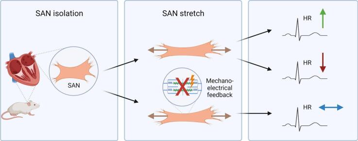
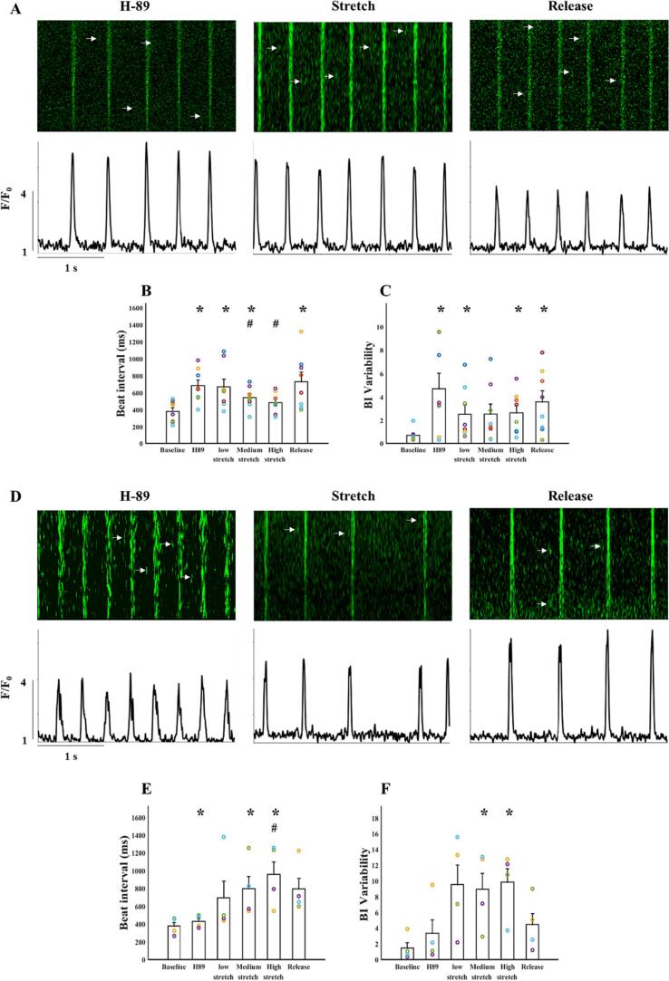
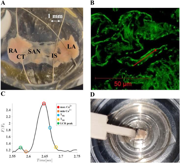
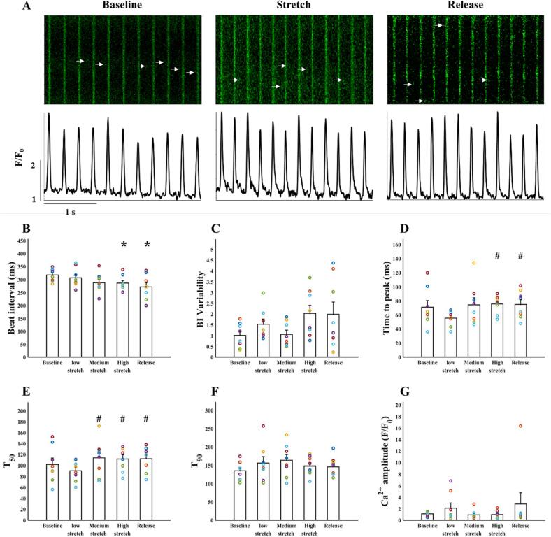
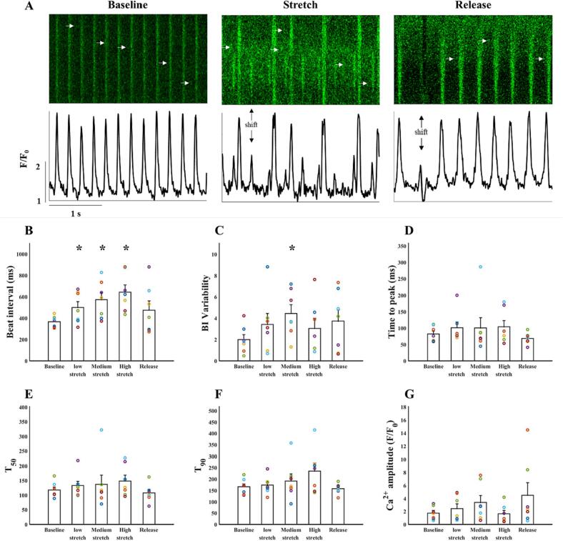
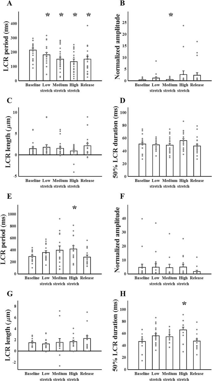
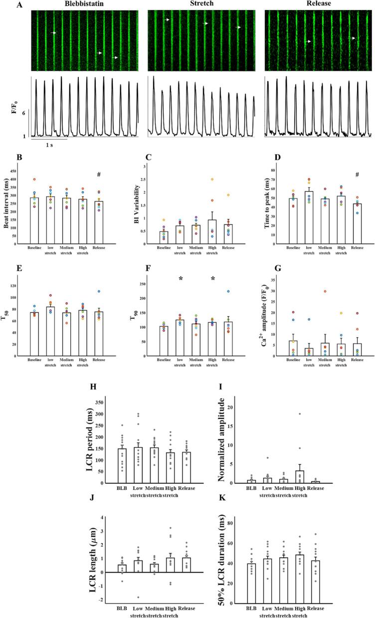
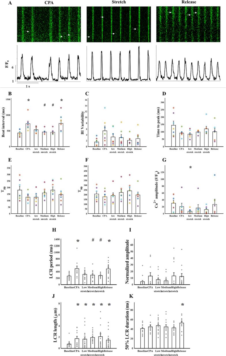
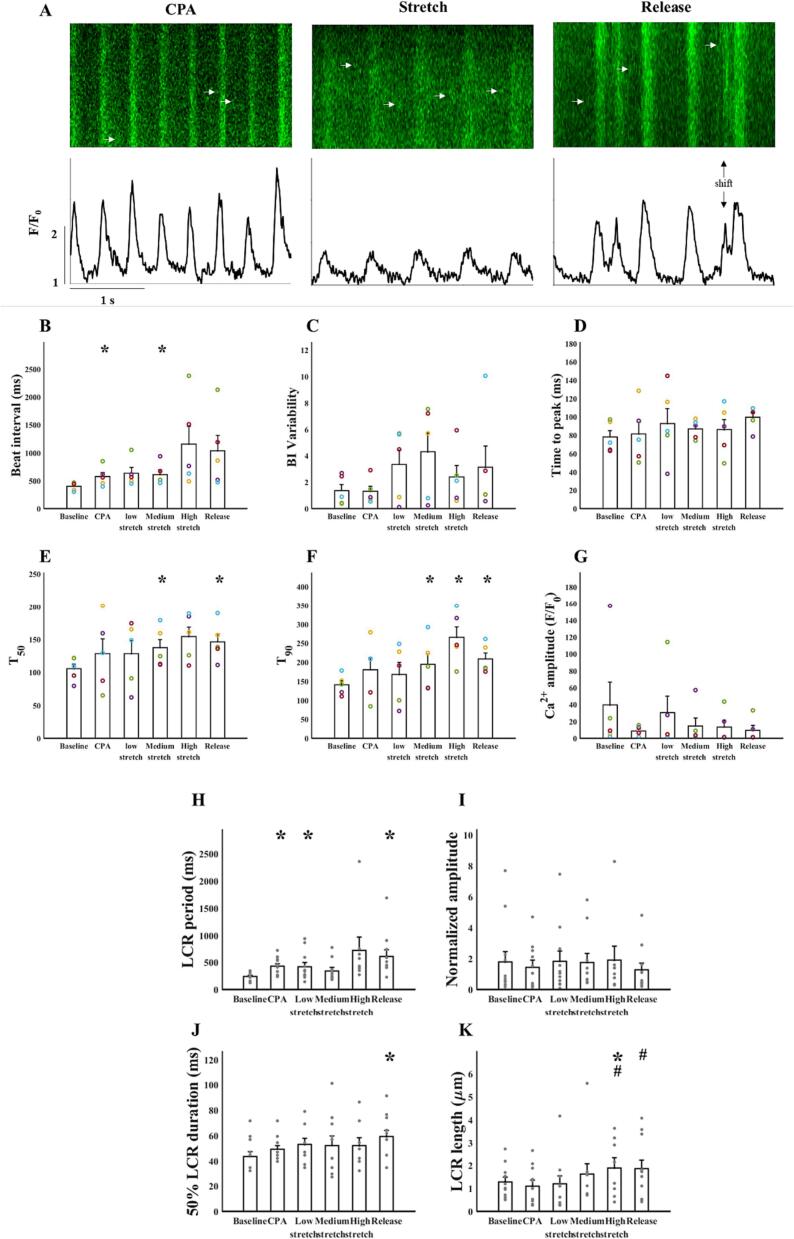
The sinoatrial node (SAN) is the primary heart pacemaker. The automaticity of SAN pacemaker cells is regulated by an integrated coupled-clock system. The beat interval (BI) of SAN, and its primary initiation location (inferior vs. superior) are determined by mutual entrainment among pacemaker cells and interaction with extrinsic effectors, including increased venous return which stretches the SAN. We aim to understand the mechanisms that link stretch to changes in BI and to heterogeneity of BI in the SAN. Isolated SAN tissues of C57BL/6 mice were gradually stretched to different degrees [(low (5-10 % lengthening), medium (10-20 %), and high (20-40 %))] using motor controlled with a custom-made Arduino software. Recordings were acquired 30 s following each level of step. In 8/15 tissues, stretch led to a positive chronotropic response, while in 7/15 tissues, a negative chronotropic response was observed. In the positive chronotropic response group, BI was shortened in parallel to shortening of the local Ca release (LCR) period, a readout of the degree of clock coupling. In the negative chronotropic response group, in parallel to a prolongation of BI and LCR period, an unsynchronized firing rate was observed among the cells upon application of stretch. Eliminating the mechano-electrical feedback by addition of blebbistatin disabled the stretch-induced chronotropic effect. Reduction of the sarcoplasmic reticulum Ca levels, which mediates the mechano-electrical feedback, by addition of cyclopiazonic acid disabled the dual effect of stretch on SAN function and BI heterogeneity. Thus, the mechano-electric feedback mediates the dual effect of stretch in mouse SAN tissue.
窦房结(SAN)是心脏的主要起搏器。SAN起搏细胞的自律性由一个整合的耦合时钟系统调节。SAN的心动周期(BI)及其主要起始位置(下部与上部)由起搏细胞之间的相互夹带以及与外部效应器的相互作用决定,外部效应器包括增加的静脉回流,它会拉伸SAN。我们旨在了解将拉伸与BI变化以及SAN中BI的异质性联系起来的机制。使用定制的Arduino软件控制的电机,将C57BL/6小鼠的离体SAN组织逐渐拉伸至不同程度[(低(延长5 - 10%)、中(10 - 20%)和高(20 - 40%))]。在每个步骤水平后30秒进行记录。在15个组织中的8个中,拉伸导致正性变时反应,而在15个组织中的7个中,观察到负性变时反应。在正性变时反应组中,BI缩短与局部钙释放(LCR)期的缩短平行,LCR期是时钟耦合程度的一个指标。在负性变时反应组中,与BI和LCR期的延长平行,在施加拉伸时细胞之间观察到不同步的放电频率。通过添加blebbistatin消除机械电反馈使拉伸诱导的变时效应失效。通过添加环匹阿尼酸降低介导机械电反馈的肌浆网钙水平,使拉伸对SAN功能和BI异质性的双重作用失效。因此,机械电反馈介导了拉伸对小鼠SAN组织的双重作用。