Chen Liping, Ye Zhenghao, Li Junhua, Wang Lijia, Chen Yu, Yu Meiping, Han Jian, Huang Jiangeng, Li Dongyan, Lv Yongling, Xiong Kai, Tian De'an, Liao Jiazhi, Seidler Ursula, Xiao Fang
Department of Gastroenterology, Tongji Hospital, Tongji Medical College, Huazhong University of Science and Technology, Wuhan, Hubei, 430030, People's Republic of China.
Department of Gastroenterology, Hannover Medical School, Hannover, Germany.
J Transl Med. 2025 Jan 13;23(1):55. doi: 10.1186/s12967-024-05873-6.
The conversion of primary bile acids to secondary bile acids by the gut microbiota has been implicated in colonic inflammation. This study investigated the role of gut microbiota related bile acid metabolism in colonic inflammation in both patients with inflammatory bowel disease (IBD) and a murine model of dextran sulfate sodium (DSS)-induced colitis.
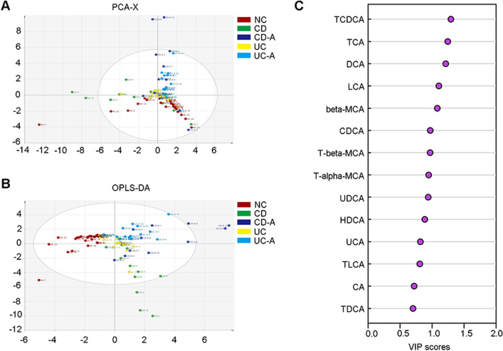
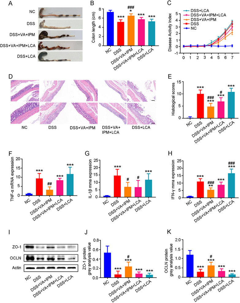
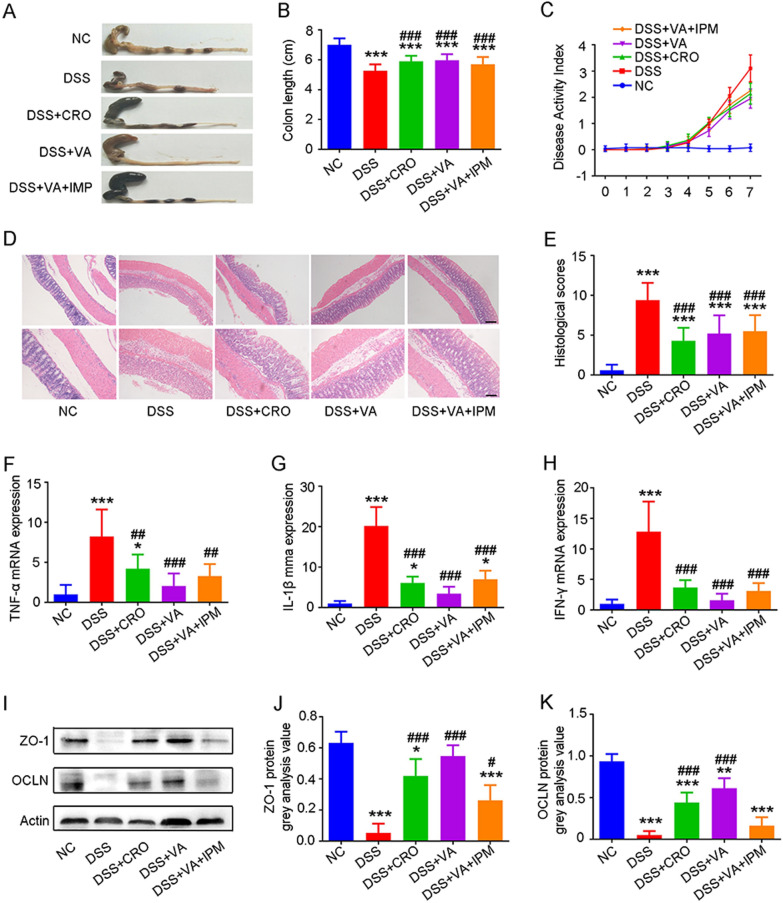
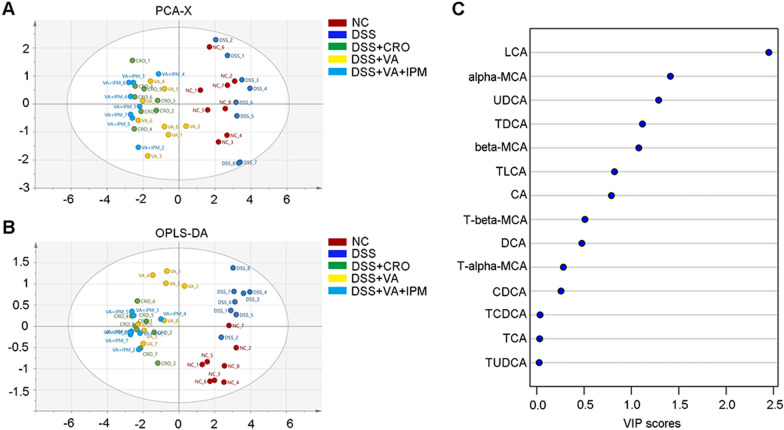
Bile acids in fecal samples from patients with IBD and DSS-induced colitis mice, with and without antibiotic treatment, were analyzed using ultraperformance liquid chromatography-mass spectrometry (UPLC-MS). The composition of the microbiota in fecal samples from IBD patients and DSS-colitis mice was characterized via Illumina MiSeq sequencing of the bacterial 16S rRNA gene V3-V4 region. Metagenomic profiling further identified metabolism-related gene signatures in stool samples from DSS-colitis mice. Histological analysis, quantitative PCR (qPCR) and Western Blotting were conducted on colonic samples from DSS-induced colitis mice to assess colonic inflammation, mucosal barrier integrity, and associated signaling pathways. The multivariate analysis of bile acids was conducted using Soft Independent Modelling of Class Analogy (SIMCA, Umetrics, Sweden). The relation between the relative abundance of specific phyla/genera and bile acid concentration was assess through Spearman's correlation analyses. Finally, lithocholic acid (LCA), the key bile acid, was administered via gavage to evaluate its effect on colonic inflammation and mucosal barrier integrity.
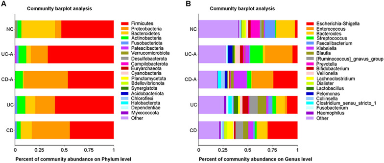
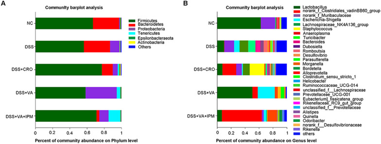
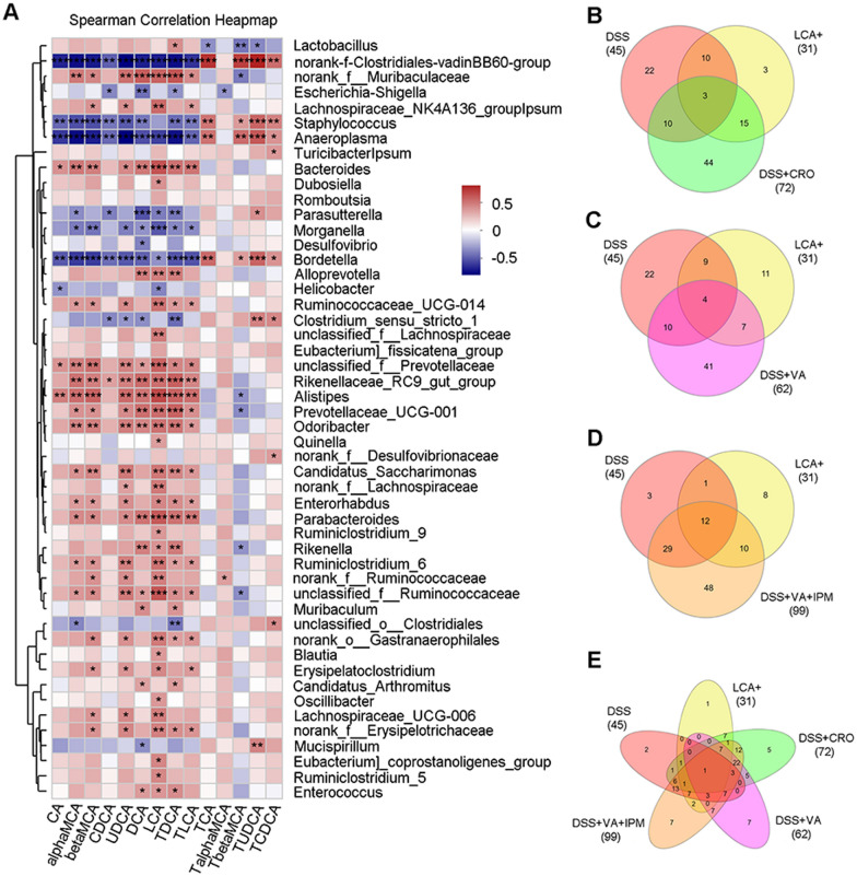
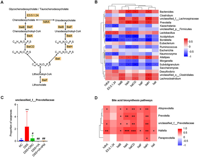
In patients with IBD, the composition of colonic bile acids and gut microbiota was altered. Moreover, changes in the gut microbiota further modulate the composition of bile acids in the intestine. As the gut microbiota continues to shift, the bile acid profile undergoes additional alterations. The aforementioned alterations were also observed in mice with DSS-induced colitis. The study revealed a correlation between dysbiosis of the gut microbiota and modifications in the profile of colonic bile acids, notably LCA observed in both patients with IBD and mice with DSS-induced colitis. Through multivariate analysis, LCA was identified as the key bile acid that significantly affects colonic inflammation and the integrity of mucosal barrier. Subsequent experiments confirmed that LCA supplementation effectively mitigated the inhibitory effects of gut microbiota on colitis progression in mice, primarily through the activation of the sphingosine-1-phosphate receptor 2 (S1PR2)/NF-κB p65 signaling pathway. Analysis of the microbiome and metagenomic data revealed changes in the gut microbiota, notably an increased abundance of an unclassified genus within the family Prevotellaceae in DSS-induced colitis mice. Furthermore, a positive correlation was observed between the relative abundance of Prevotellaceae and bile acid biosynthesis pathways, as well as colonic LCA level.
These findings suggest that LCA and its positively correlated gut bacteria, Prevotellaceae, are closely associated with intestinal inflammation. Targeting colonic inflammation may involve inhibiting LCA and members of the Prevotellaceae family as potential therapeutic strategies.
肠道微生物群将初级胆汁酸转化为次级胆汁酸与结肠炎症有关。本研究调查了肠道微生物群相关胆汁酸代谢在炎症性肠病(IBD)患者和葡聚糖硫酸钠(DSS)诱导的结肠炎小鼠模型的结肠炎症中的作用。
使用超高效液相色谱-质谱联用仪(UPLC-MS)分析IBD患者和DSS诱导的结肠炎小鼠粪便样本中有无抗生素治疗情况下的胆汁酸。通过对细菌16S rRNA基因V3-V4区域进行Illumina MiSeq测序,对IBD患者和DSS结肠炎小鼠粪便样本中的微生物群组成进行表征。宏基因组分析进一步确定了DSS结肠炎小鼠粪便样本中与代谢相关的基因特征。对DSS诱导的结肠炎小鼠的结肠样本进行组织学分析、定量PCR(qPCR)和蛋白质免疫印迹,以评估结肠炎症、粘膜屏障完整性及相关信号通路。使用类分析的软独立建模(SIMCA,瑞典Umetrics公司)对胆汁酸进行多变量分析。通过Spearman相关性分析评估特定门/属的相对丰度与胆汁酸浓度之间的关系。最后,通过灌胃给予关键胆汁酸石胆酸(LCA),以评估其对结肠炎症和粘膜屏障完整性的影响。
IBD患者的结肠胆汁酸和肠道微生物群组成发生了改变。此外,肠道微生物群的变化进一步调节了肠道中胆汁酸的组成。随着肠道微生物群持续变化,胆汁酸谱会发生更多改变。在DSS诱导的结肠炎小鼠中也观察到上述变化。该研究揭示了肠道微生物群失调与结肠胆汁酸谱改变之间的相关性,特别是在IBD患者和DSS诱导的结肠炎小鼠中均观察到的LCA。通过多变量分析,LCA被确定为显著影响结肠炎症和粘膜屏障完整性的关键胆汁酸。随后的实验证实,补充LCA可有效减轻肠道微生物群对小鼠结肠炎进展的抑制作用,主要是通过激活鞘氨醇-1-磷酸受体2(S1PR2)/NF-κB p65信号通路。对微生物组和宏基因组数据的分析揭示了肠道微生物群的变化,特别是在DSS诱导的结肠炎小鼠中,普雷沃氏菌科内一个未分类属的丰度增加。此外,观察到普雷沃氏菌科的相对丰度与胆汁酸生物合成途径以及结肠LCA水平之间呈正相关。
这些发现表明,LCA及其正相关的肠道细菌普雷沃氏菌科与肠道炎症密切相关。针对结肠炎症的治疗可能涉及抑制LCA和普雷沃氏菌科成员,作为潜在的治疗策略。