Rosenblum Jared S, Cole Yasemin, Dang Danielle, Lookian Pashayar P, Alkaissi Hussam, Patel Mayank, Cappadona Anthony J, Jha Abhishek, Edwards Nancy, Donahue Danielle R, Munasinghe Jeeva, Wang Herui, Knutsen Russell H, Pappo Alberto S, Lechan Ronald M, Kozel Beth A, Smirniotopoulos James G, Kim H Jeffrey, Vortmeyer Alexander, Miettinen Markku, Heiss John D, Zhuang Zhengping, Pacak Karel
Neuro-Oncology Branch, National Cancer Institute, National Institutes of Health, Bethesda, MD, United States.
National Institute of Diabetes and Digestive and Kidney Diseases, National Institutes of Health, Bethesda, MD, United States.
JNCI Cancer Spectr. 2025 Jan 3;9(1). doi: 10.1093/jncics/pkaf001.
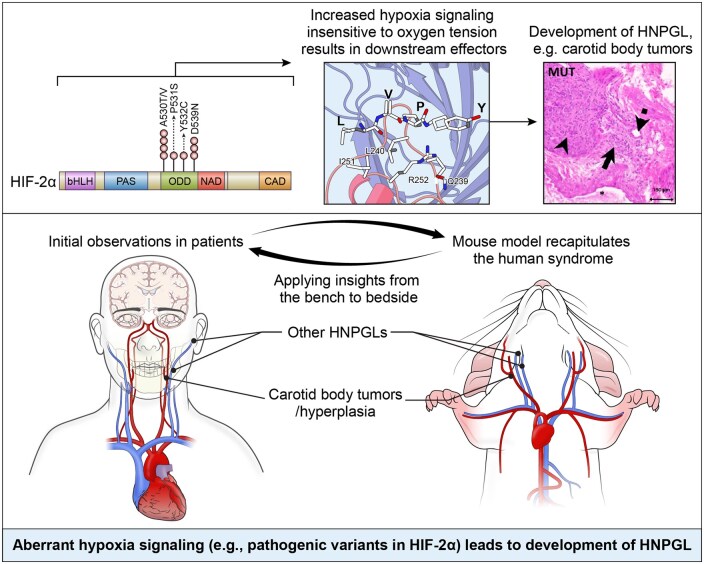
Head and neck paragangliomas (HNPGLs) are typically slow-growing, hormonally inactive tumors of parasympathetic paraganglia. Inactivation of prolyl-hydroxylase domain-containing 2 protein causing indirect gain-of-function of hypoxia-inducible factor-2α (HIF-2α), encoded by EPAS1, was recently shown to cause carotid body hyperplasia. We previously described a syndrome with multiple sympathetic paragangliomas caused by direct gain-of-function variants in EPAS1 (Pacak-Zhuang syndrome, PZS) and developed a corresponding mouse model.
We evaluated a cohort of patients with PZS (n = 9) for HNPGL by positron emission tomography, magnetic resonance imaging, and computed tomography and measured carotid body size compared to literature reference values. Resected tumors were evaluated by histologic sectioning and staining. We evaluated the corresponding mouse model at multiple developmental stages (P8 and adult) for lesions of the head and neck by high resolution ex vivo imaging and performed immunohistochemical staining on histologic sections of the identified lesions.
hree patients had imaging consistent with HNPGL, one of which warranted resection and was confirmed on histology. Three additional patients had carotid body enlargement (Z-score > 2.0), and 3 had carotid artery malformations. We found that 9 of 10 adult variant mice had carotid body tumors and 6 of 8 had a paraganglioma on the cranio-caval vein, the murine homologue of the superior vena cava; these were also found in 4 of 5 variant mice at post-natal day 8. These tumors and the one resected from a patient were positive for tyrosine hydroxylase, synaptophysin, and chromogranin A. Brown fat in a resected patient tumor carried the EPAS1 pathogenic variant.
These findings (1) suggest HNPGL as a feature of PZS and (2) show that these pathogenic variants are sufficient to cause the development of these tumors, which we believe represents a continuous spectrum of disease starting from hyperplasia.
头颈部副神经节瘤(HNPGLs)通常是生长缓慢、无激素活性的副交感神经节肿瘤。最近研究表明,含脯氨酰羟化酶结构域的2蛋白失活导致缺氧诱导因子-2α(HIF-2α,由EPAS1编码)间接功能获得,可引起颈动脉体增生。我们之前描述了一种由EPAS1中直接功能获得性变异导致的多发性交感神经节瘤综合征(Pacak-Zhuang综合征,PZS),并建立了相应的小鼠模型。
我们通过正电子发射断层扫描、磁共振成像和计算机断层扫描对一组PZS患者(n = 9)进行HNPGL评估,并将颈动脉体大小与文献参考值进行比较。对切除的肿瘤进行组织切片和染色评估。我们通过高分辨率离体成像在多个发育阶段(出生后第8天和成年期)评估相应的小鼠模型的头颈部病变,并对所识别病变的组织切片进行免疫组织化学染色。
3例患者的影像学检查结果与HNPGL一致,其中1例需要切除,组织学检查得以证实。另外3例患者有颈动脉体增大(Z评分>2.0),3例有颈动脉畸形。我们发现,10只成年变异小鼠中有9只有颈动脉体肿瘤,8只中有6只在颅腔静脉(相当于人类上腔静脉的小鼠同源物)上有副神经节瘤;在出生后第8天的5只变异小鼠中,有4只也发现了这些肿瘤。这些肿瘤以及从一名患者身上切除的肿瘤对酪氨酸羟化酶、突触素和嗜铬粒蛋白A呈阳性。一名患者切除肿瘤中的棕色脂肪携带EPAS1致病变异。
这些发现(1)表明HNPGL是PZS的一个特征,(2)表明这些致病变异足以导致这些肿瘤的发生,我们认为这代表了从增生开始的连续疾病谱。