Soeratanapant Suphat, Sutthigran Somchin, Saisawart Phasamon, Chaivoravitsakul Nardtiwa, Horoongruang Kongthit, Limpongsai Luksamee, Tantarawanich Artima, Thanaboonnipat Chutimon, Tachampa Kittipong, Choisunirachon Nan
Department of Surgery, Faculty of Veterinary Science, Chulalongkorn University, Bangkok, Thailand.
The Small Animal Hospital, Faculty of Veterinary Science, Small Animal Teaching Hospital, Chulalongkorn University, Bangkok, Thailand.
Vet World. 2024 Nov;17(11):2635-2643. doi: 10.14202/vetworld.2024.2635-2643. Epub 2024 Nov 25.
Computed tomographic (CT) images can elucidate the variations of cardiac orientation that this information among dog breeds has never been reported. This study aimed to explore the heart orientations of dogs with different thoracic types and study their effects on vertebral heart score (VHS) measurements using CT images.
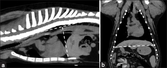
Thoracic CT images of 115 mature dogs without thoracic abnormalities were retrospectively examined. The dogs were classified into four groups: Normal Broad, Abnormal Broad, Normal, and Deep. All dogs were also classified based on their heart deviations. The VHSs were evaluated using lateral VHS, dorsal VHS, and adjusted VHS, and all were compared.
In the normal broad and abnormal broad groups, the lateral VHS and lateral long-axis dimensions were significantly lower than those obtained from the dorsal and adjusted VHSs. In addition, heart deviations were mostly observed in the normal broad and abnormal broad groups. Nevertheless, little evidence was found in the normal and deep groups. The lateral VHS and lateral long-axis dimensions were significantly reduced by heart deviation more than the dorsal and adjusted VHSs.
Cardiac orientations among dog breeds can affect VHSs of lateral projection, especially in the broad thoracic group. Clinical evaluation of the VHS in the broad thoracic dogs should be performed on the dorsal view for more accurate measurement of heart size.
计算机断层扫描(CT)图像能够阐明心脏方位的变化,但不同犬种间的此类信息此前从未有过报道。本研究旨在利用CT图像探究不同胸廓类型犬的心脏方位,并研究其对心脏椎体评分(VHS)测量的影响。
回顾性检查了115只无胸廓异常的成年犬的胸部CT图像。这些犬被分为四组:正常宽胸型、异常宽胸型、正常型和深胸型。所有犬还根据其心脏偏移情况进行了分类。使用侧位VHS、背位VHS和校正后的VHS评估VHS,并对所有评估结果进行比较。
在正常宽胸型和异常宽胸型组中,侧位VHS和侧位长轴尺寸显著低于背位和校正后的VHS所测得的值。此外,心脏偏移大多见于正常宽胸型和异常宽胸型组。然而,在正常型和深胸型组中未发现明显证据。与背位和校正后的VHS相比,心脏偏移对侧位VHS和侧位长轴尺寸的减小影响更大。
不同犬种间的心脏方位会影响侧位投影的VHS,尤其是在宽胸型犬组中。对于宽胸型犬,应在背位视图上对VHS进行临床评估,以更准确地测量心脏大小。