Maximova Olga A, Anzick Sarah L, Sturdevant Daniel E, Bennett Richard S, Faucette Lawrence J, St Claire Marisa, Whitehead Stephen S, Kanakabandi Kishore, Sheng Zong-Mei, Xiao Yongli, Kash John C, Taubenberger Jeffery K, Martens Craig, Cohen Jeffrey I
Laboratory of Infectious Diseases, National Institute of Allergy and Infectious Diseases, National Institutes of Health; Bethesda, Maryland, United States of America.
Rocky Mountain Laboratories, Research Technologies Branch, Genomics Research Section, National Institute of Allergy and Infectious Diseases, National Institutes of Health; Hamilton, Montana, United States of America.
PLoS Pathog. 2025 Jan 22;21(1):e1012530. doi: 10.1371/journal.ppat.1012530. eCollection 2025 Jan.
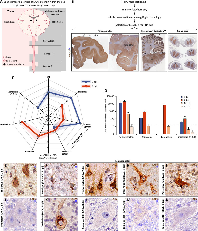
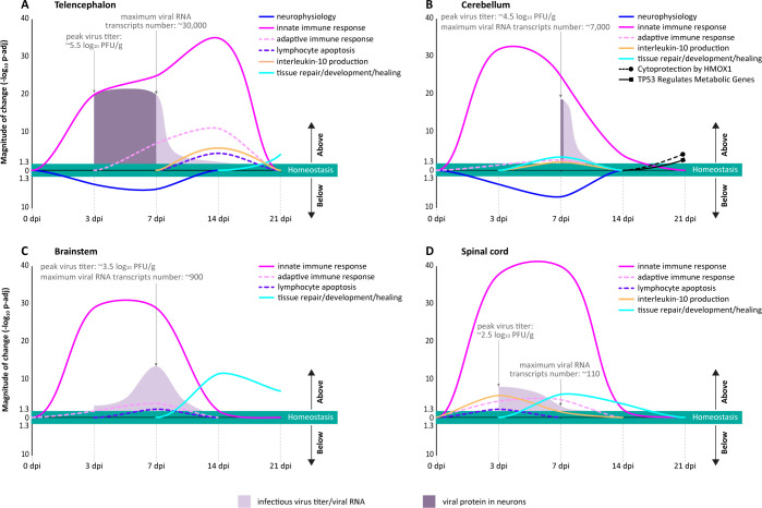
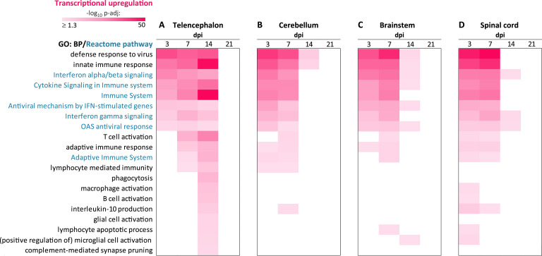
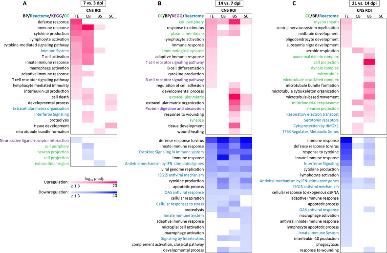
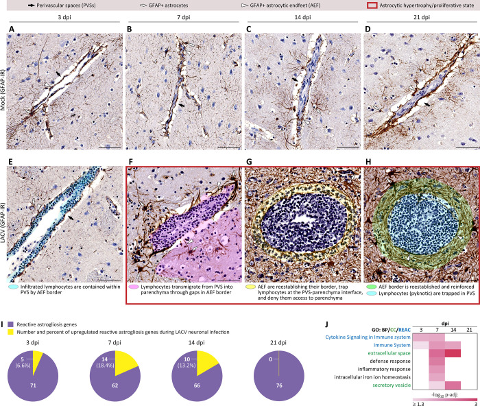
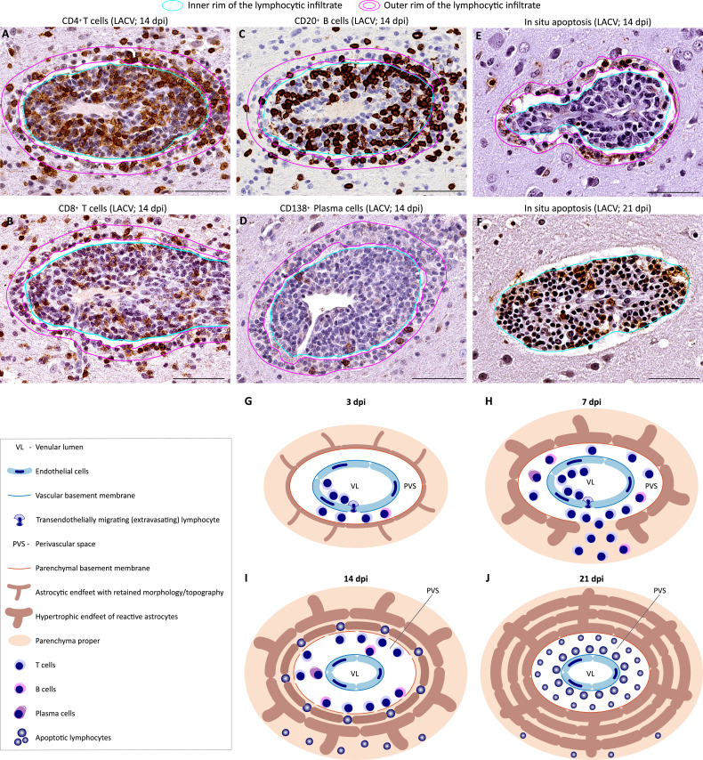
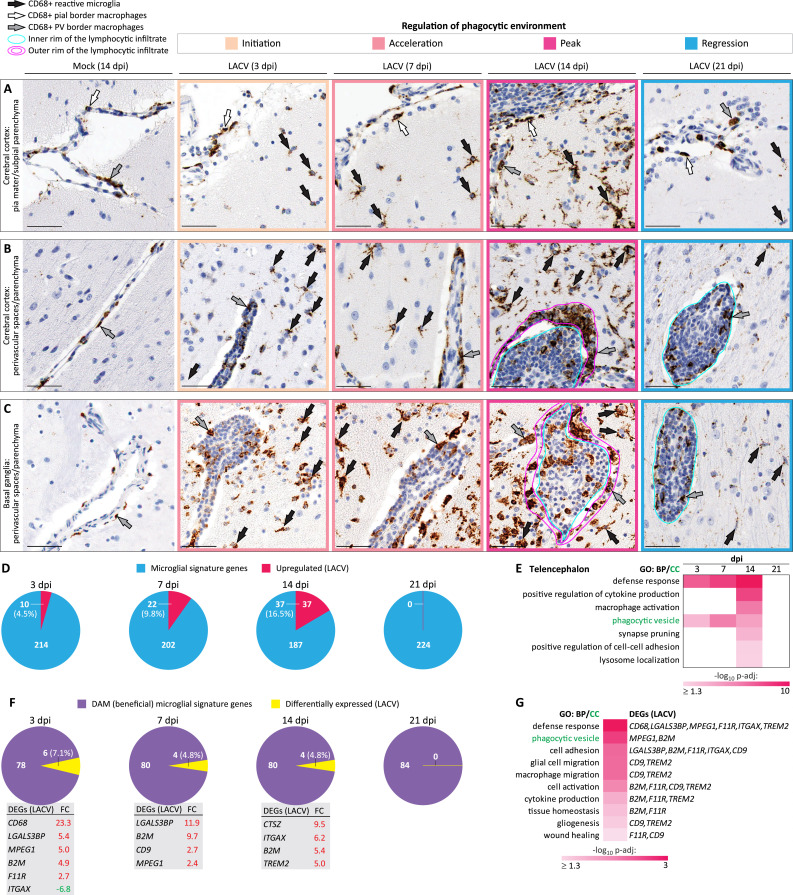
Viral infections of the central nervous system (CNS) are a major cause of morbidity largely due to lack of prevention and inadequate treatments. While mortality from viral CNS infections is significant, nearly two thirds of the patients survive. Thus, it is important to understand how the human CNS can successfully control virus infection and recover. Since it is not possible to study the human CNS throughout the course of viral infection at the cellular level, here we analyzed a non-lethal viral infection in the CNS of nonhuman primates (NHPs). We inoculated NHPs intracerebrally with a high dose of La Crosse virus (LACV), a bunyavirus that can infect neurons and cause encephalitis primarily in children, but with a very low (≤ 1%) mortality rate. To profile the CNS response to LACV infection, we used an integrative approach that was based on comprehensive analyses of (i) spatiotemporal dynamics of virus replication, (ii) identification of types of infected neurons, (iii) spatiotemporal transcriptomics, and (iv) morphological and functional changes in CNS intrinsic and extrinsic cells. We identified the location, timing, and functional repertoire of optimal transcriptional and translational regulation of the primate CNS in response to virus infection of neurons. These CNS responses involved a well-coordinated spatiotemporal interplay between astrocytes, lymphocytes, microglia, and CNS-border macrophages. Our findings suggest a multifaceted program governing an optimal CNS response to virus infection with specific events coordinated in space and time. This allowed the CNS to successfully control the infection by rapidly clearing the virus from infected neurons, mitigate damage to neurophysiology, activate and terminate immune responses in a timely manner, resolve inflammation, restore homeostasis, and initiate tissue repair. An increased understanding of these processes may provide new therapeutic opportunities to improve outcomes of viral CNS diseases in humans.
中枢神经系统(CNS)的病毒感染是发病的主要原因,这在很大程度上是由于缺乏预防措施和治疗方法不足。虽然病毒性中枢神经系统感染的死亡率很高,但近三分之二的患者能够存活。因此,了解人类中枢神经系统如何成功控制病毒感染并恢复健康非常重要。由于不可能在细胞水平上研究人类中枢神经系统在病毒感染全过程中的情况,我们在此分析了非人灵长类动物(NHP)中枢神经系统中的非致死性病毒感染。我们向NHP脑内接种了高剂量的拉克罗斯病毒(LACV),这是一种布尼亚病毒,可感染神经元并主要导致儿童脑炎,但其死亡率非常低(≤1%)。为了描述中枢神经系统对LACV感染的反应,我们采用了一种综合方法,该方法基于对以下方面的全面分析:(i)病毒复制的时空动态,(ii)被感染神经元类型的鉴定,(iii)时空转录组学,以及(iv)中枢神经系统内在和外在细胞的形态和功能变化。我们确定了灵长类中枢神经系统在神经元病毒感染时最佳转录和翻译调控的位置、时间和功能范围。这些中枢神经系统反应涉及星形胶质细胞、淋巴细胞、小胶质细胞和中枢神经系统边界巨噬细胞之间协调良好的时空相互作用。我们的研究结果表明,一个多方面的程序控制着中枢神经系统对病毒感染的最佳反应,特定事件在空间和时间上得到协调。这使得中枢神经系统能够通过迅速从受感染神经元中清除病毒来成功控制感染,减轻对神经生理学的损害,及时激活和终止免疫反应,解决炎症,恢复体内平衡,并启动组织修复。对这些过程的进一步了解可能为改善人类病毒性中枢神经系统疾病的治疗效果提供新的治疗机会。