Luna-Gutiérrez Myrna, Azorín-Vega Erika, Oros-Pantoja Rigoberto, Ocampo-García Blanca, Cruz-Nova Pedro, Jiménez-Mancilla Nallely, Bravo-Villegas Gerardo, Santos-Cuevas Clara, Meléndez-Alafort Laura, Ferro-Flores Guillermina
Department of Radioactive Materials, Instituto Nacional de Investigaciones Nucleares, Ocoyoacac, 52750, Mexico.
Faculty of Medicine, Universidad Autónoma del Estado de México, Toluca, 50180, Mexico.
EJNMMI Radiopharm Chem. 2025 Jan 22;10(1):5. doi: 10.1186/s41181-025-00328-9.
Cancer immunotherapy is a relatively new approach to cancer treatment. Peptides that target specific pathways and cells involved in immunomodulation can potentially improve the efficacy of cancer therapy. Recently, we reported iPD-L1 as a novel inhibitor peptide that specifically targets the cancer cell ligand PD-L1 (programmed death ligand 1). PD-L1 is responsible for inhibiting the immune checkpoint protein PD-1 expressed by regulatory T cells. On the other hand, anti-PD-L1 immunotherapy in combination with external beam radiotherapy has shown improved outcomes in the treatment of breast and lung cancer. The aim of this research was to prepare Lu-labeled iPD-L1 and to preclinically evaluate its radiotherapeutic potential and role as a tumor immunomodulator by measuring macrophage activation, IL-10, TGFβ, and PD-L1 expression in 4T1 triple-negative breast cancer cells and murine 4T1 tumors after treatment with Lu-iPD-L1.
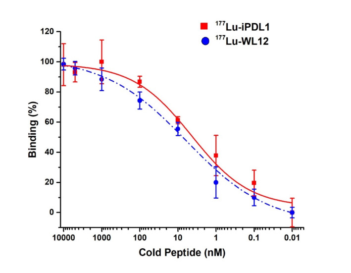
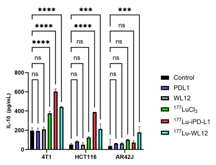
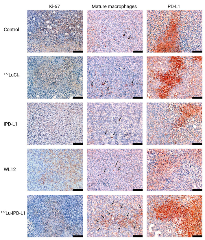
The iPD-L1 ligand, characterized by UPLC mass, UV-Vis, and FT-IR spectroscopies, showed a chemical purity of 99%. The Lu-iPD-L1 radiochemical purity was 98.9 ± 1.1%. In vitro and in vivo studies demonstrated radiotracer stability in human serum (> 97% after 24 h evaluated by radio-HPLC), adequate affinity by the PDL1 protein (IC = 4.21 nM), and specific detection for PD-L1 assessed in 4T1, HCT116, and AR42J cancer cells, in which PD-L1 expression was verified by immunofluorescence and Western Blot assays. After treatment with Lu-iPD-L1 (0.4 Bq/cell), flow cytometry results showed a significant decrease in cell viability of 4T1 cells (dead 56.2%) compared to LuCl (dead 34.2%) and untreated cells (dead 9.4%). With high tumor uptake (6.97 ± 1.04%ID) and hepatobiliary and renal clearance, lutetium-177-labeled iPD-L1 delivered a tumor dose of 27 Gy/37 MBq and less than 0.36 Gy/37 MBq to non-source organs. PD-L1 positive tumors showed a significant increase in activated macrophages, PD-L1, IL-10, and TGFβ expression levels after Lu-iPD-L1 treatment as evaluated by ELISA assay and immunohistochemistry.
Therefore, this study warrants further dosimetric and clinical studies to determine the immunomodulatory effect and therapeutic efficacy of Lu-iPD-L1 in treating PD-L1-positive tumors in combination with anti-PD-1/PD-L1 immunotherapy protocols.
癌症免疫疗法是一种相对较新的癌症治疗方法。靶向参与免疫调节的特定途径和细胞的肽可能会提高癌症治疗的疗效。最近,我们报道了iPD-L1作为一种新型抑制剂肽,它特异性靶向癌细胞配体PD-L1(程序性死亡配体1)。PD-L1负责抑制调节性T细胞表达的免疫检查点蛋白PD-1。另一方面,抗PD-L1免疫疗法与外照射放疗联合应用已显示出在乳腺癌和肺癌治疗中改善了疗效。本研究的目的是制备镥标记的iPD-L1,并通过测量用镥-iPD-L1处理后的4T1三阴性乳腺癌细胞和小鼠4T1肿瘤中的巨噬细胞活化、IL-10、TGFβ和PD-L1表达,对其放射治疗潜力和作为肿瘤免疫调节剂的作用进行临床前评估。
通过超高效液相色谱质谱、紫外可见光谱和傅里叶变换红外光谱表征的iPD-L1配体,化学纯度为99%。镥-iPD-L1的放射化学纯度为98.9±1.1%。体外和体内研究表明,放射性示踪剂在人血清中具有稳定性(通过放射性高效液相色谱法评估,24小时后>97%),对PDL1蛋白具有足够的亲和力(IC = 4.21 nM),并且在4T1、HCT116和AR42J癌细胞中对PD-L1有特异性检测,其中通过免疫荧光和蛋白质印迹分析验证了PD-L1的表达。在用镥-iPD-L1(0.4 Bq/细胞)处理后,流式细胞术结果显示,与氯化镥(死亡34.2%)和未处理细胞(死亡9.4%)相比,4T1细胞的活力显著降低(死亡56.2%)。镥-177标记的iPD-L1具有高肿瘤摄取(6.97±1.04%ID)以及肝胆和肾脏清除,给肿瘤的剂量为27 Gy/37 MBq,给非靶器官的剂量小于0.36 Gy/37 MBq。通过酶联免疫吸附测定和免疫组织化学评估,PD-L1阳性肿瘤在用镥-iPD-L1治疗后,活化巨噬细胞、PD-L1、IL-10和TGFβ表达水平显著增加。
因此,本研究值得进一步进行剂量学和临床研究,以确定镥-iPD-L1与抗PD-1/PD-L1免疫治疗方案联合治疗PD-L1阳性肿瘤的免疫调节作用和治疗效果。