Schlögl Kerstin, Güth Jan-Frederik, Graf Tobias, Keul Christine
Department of Prosthetic Dentistry, LMU University Hospital, LMU Munich, Goethestrasse 70, 80336, Munich, Germany.
Department of Prosthetic Dentistry, Center for Dentistry and Oral Medicine (Carolinum), Goethe University Frankfurt am Main, Frankfurt am Main, Germany.
Clin Oral Investig. 2025 Jan 27;29(1):92. doi: 10.1007/s00784-025-06154-2.
Evaluation of the accuracy of direct digitization of maxillary scans depending on the scanning strategy.
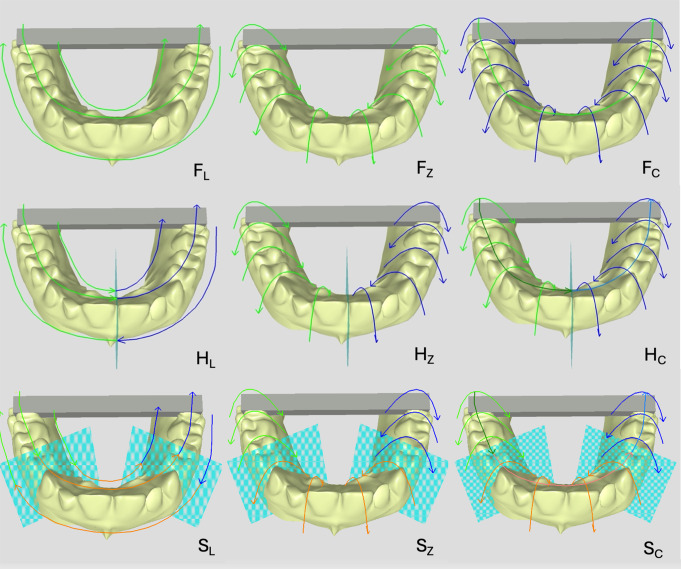
A maxillary model with a metal bar as a reference structure fixed between the second molars was digitized using the CEREC Primescan AC scanner (N = 225 scans). Nine scanning strategies were selected (n = 25 scans per strategy), differing in scan area segmentation (F = full jaw, H = half jaw, S = sextant) and scan movement pattern (L = linear, Z = zig-zag, C = combined). Trueness was assessed by evaluating linear differences in the X, Y, and Z axes and angular deviations (α axial, α coronal, α total) compared to a reference dataset. Statistical differences were analyzed using Kruskal-Wallis and Mann-Whitney U tests (p<0.017). Precision was analyzed by the standard deviation of linear and angular aberrations (ISO 5725-1) (p < 0.05).
Strategy F showed significantly higher trueness and precision than F for VE (p = 0.009), V(y) (p = 0.010), α (p = 0.004), and α (p = 0.002). Strategy F demonstrated significantly better trueness than F for VE (p = 0.007), α (p = 0.010), and α (p = 0.013). For scan segmentation, F showed better trueness for V(y) (p = 0.001) and α (p < 0.001) than H. Strategy H showed better trueness for V(z) than for F and S (p = 0.001, p = 0.002). The scanning patterns F, F, and H exhibited the best performance for trueness and precision.
Scanning motion and segmentation have a significant impact on the trueness and precision of full-arch scans.
The scanning strategy is decisive in enhancing the clinical workflow and the accuracy of full-arch scans.
根据扫描策略评估上颌扫描直接数字化的准确性。
使用CEREC Primescan AC扫描仪对上颌模型进行数字化,该模型在第二磨牙之间固定有一根金属棒作为参考结构(N = 225次扫描)。选择了九种扫描策略(每种策略n = 25次扫描),在扫描区域分割(F = 全颌,H = 半颌,S = 牙弓段)和扫描移动模式(L = 线性,Z = 之字形,C = 组合)方面存在差异。通过评估与参考数据集相比X、Y和Z轴上的线性差异以及角度偏差(α轴向、α冠状、α总和)来评估准确性。使用Kruskal-Wallis和Mann-Whitney U检验分析统计差异(p<0.017)。通过线性和角度像差的标准偏差(ISO 5725-1)分析精度(p < 0.05)。
对于VE(p = 0.009)、V(y)(p = 0.010)、α(p = 0.004)和α(p = 0.002),策略F显示出比F显著更高的准确性和精度。对于VE(p = 0.007)、α(p = 0.010)和α(p = 0.013),策略F显示出比F显著更好的准确性。对于扫描分割,F在V(y)(p = 0.001)和α(p < 0.001)方面显示出比H更好的准确性。策略H在V(z)方面显示出比F和S更好的准确性(p = 0.001,p = 0.002)。扫描模式F、F和H在准确性和精度方面表现最佳。
扫描运动和分割对全牙弓扫描的准确性和精度有显著影响。
扫描策略对于优化临床工作流程和全牙弓扫描的准确性起着决定性作用。