• 文献检索
  • 文档翻译
  • 深度研究
  • 学术资讯
  • Suppr Zotero 插件Zotero 插件
  • 邀请有礼
  • 套餐&价格
  • 历史记录
应用&插件
Suppr Zotero 插件Zotero 插件浏览器插件Mac 客户端Windows 客户端微信小程序
定价
高级版会员购买积分包购买API积分包
服务
文献检索文档翻译深度研究API 文档MCP 服务
关于我们
关于 Suppr公司介绍联系我们用户协议隐私条款
关注我们

Suppr 超能文献

核心技术专利:CN118964589B侵权必究
粤ICP备2023148730 号-1Suppr @ 2026

文献检索

告别复杂PubMed语法,用中文像聊天一样搜索,搜遍4000万医学文献。AI智能推荐,让科研检索更轻松。

立即免费搜索

文件翻译

保留排版,准确专业,支持PDF/Word/PPT等文件格式,支持 12+语言互译。

免费翻译文档

深度研究

AI帮你快速写综述,25分钟生成高质量综述,智能提取关键信息,辅助科研写作。

立即免费体验

在有峡部的根管中使用不同封闭剂放置方法的单锥充填技术后空隙的存在及分布情况。

Presence and distribution of voids after using the single cone obturation technique with different sealer placement methods in canals with an isthmus.

作者信息

Ma Yu-Qi, Liang Yu-Hong

机构信息

Department of Cariology and Endodontology, Peking University School and Hospital of Stomatology & National Center for Stomatology & National Clinical Research Center for Oral Diseases & National Engineering Research Center of Oral Biomaterials and Digital Medical Devices& Beijing Key Laboratory of Digital Stomatology & NHC Key Laboratory of Digital Stomatology & NMPA Key Laboratory for Dental Materials, Beijing, China.

Department of Stomatology, Peking University International Hospital, Beijing, China.

出版信息

J Dent Sci. 2025 Jan;20(1):286-291. doi: 10.1016/j.jds.2024.07.014. Epub 2024 Jul 25.

DOI:10.1016/j.jds.2024.07.014
PMID:39873083
原文链接:https://pmc.ncbi.nlm.nih.gov/articles/PMC11763567/
Abstract

BACKGROUND/PURPOSE: The obturation of canals with irregular structures is still a challenge for single cone obturation technique (SC). The purpose of this study was to evaluate the presence and distribution of voids using SC with different sealer placement methods in the canal with a simulated band-shaped isthmus.

MATERIALS AND METHODS

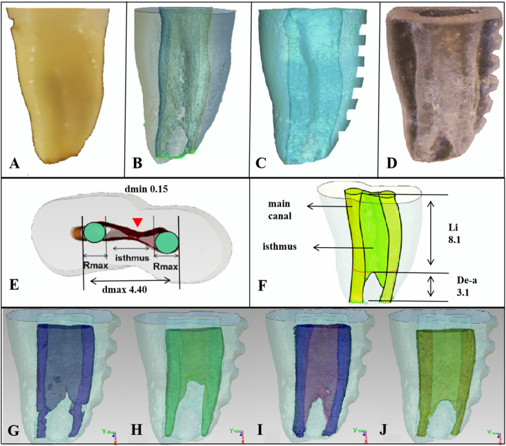
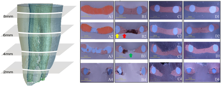
3D-printed root canal models with band-shaped isthmuses were randomly divided into four groups according to different obturation methods. Group 1: sealer placement by single cone passively (SCP); Group 2: bi-directional spiral-supported sealer placement (BS); Group 3: ultrasound-supported sealer placement (U). Group 4: vertical compaction obturation (VC). In each group, 10 of 14 models were sliced and the remaining four were scanned by micro-CT. The percentage area of voids (PAV) and the percentage volume of voids (PVV) of fillings were calculated.

RESULTS

At all slice levels, using BS and U to support sealer placement reduced voids with an average PAV of 21%, and in the VC and SCP groups were 33% and 45% respectively. Based on the micro-CT scans, nearly half of the porosity decreased by the BS and ultrasound in the isthmus with PVV of 25% and 29% respectively, compared with 46% in the SCP group. However, in the main canal, when the PVV was 22% in the SCP group, the porosity decreased to 14% in the U group and 18% in the BS group.

CONCLUSION

Bi-directional spiral or ultrasound-supported sealer placement can improve the performance of single cone obturation in canals with an isthmus.

摘要

背景/目的:对于单锥充填技术(SC)而言,充填具有不规则结构的根管仍是一项挑战。本研究的目的是使用不同的封闭剂放置方法,通过单锥技术评估模拟带状峡部根管内空隙的存在情况及分布。

材料与方法

将具有带状峡部的3D打印根管模型根据不同的充填方法随机分为四组。第1组:单锥被动放置封闭剂(SCP);第2组:双向螺旋辅助封闭剂放置(BS);第3组:超声辅助封闭剂放置(U)。第4组:垂直加压充填(VC)。每组中,14个模型中的10个被切片,其余4个通过显微CT扫描。计算充填物的空隙面积百分比(PAV)和空隙体积百分比(PVV)。

结果

在所有切片水平,使用BS和U辅助封闭剂放置可减少空隙,平均PAV为21%,而VC组和SCP组分别为33%和45%。基于显微CT扫描,在峡部,BS组和超声组的孔隙率分别降低了近一半,PVV分别为25%和29%,而SCP组为46%。然而,在主根管中,当SCP组的PVV为22%时,U组的孔隙率降至14%,BS组降至18%。

结论

双向螺旋或超声辅助封闭剂放置可提高单锥技术在有峡部根管中的充填性能。

https://cdn.ncbi.nlm.nih.gov/pmc/blobs/b68a/11763567/3927097f87d2/gr2.jpg
https://cdn.ncbi.nlm.nih.gov/pmc/blobs/b68a/11763567/d448e331e10c/gr1.jpg
https://cdn.ncbi.nlm.nih.gov/pmc/blobs/b68a/11763567/3927097f87d2/gr2.jpg
https://cdn.ncbi.nlm.nih.gov/pmc/blobs/b68a/11763567/d448e331e10c/gr1.jpg
https://cdn.ncbi.nlm.nih.gov/pmc/blobs/b68a/11763567/3927097f87d2/gr2.jpg

相似文献

1
Presence and distribution of voids after using the single cone obturation technique with different sealer placement methods in canals with an isthmus.在有峡部的根管中使用不同封闭剂放置方法的单锥充填技术后空隙的存在及分布情况。
J Dent Sci. 2025 Jan;20(1):286-291. doi: 10.1016/j.jds.2024.07.014. Epub 2024 Jul 25.
2
[ study of using single cone obturation technique in artificial canals with an isthmus].[单锥充填技术在带峡部人工根管中的应用研究]
Beijing Da Xue Xue Bao Yi Xue Ban. 2025 Apr 18;57(2):369-375. doi: 10.19723/j.issn.1671-167X.2025.02.023.
3
Micro-computed tomographic evaluation on the quality of single-cone obturation using a modified passive-deflation sealer injection needle: an in vitro study.使用改良被动泄气式封管注射针评估单牙胶尖根管充填质量的微计算机断层扫描:一项体外研究。
BMC Oral Health. 2024 Apr 20;24(1):476. doi: 10.1186/s12903-024-04232-7.
4
Evaluation of the Impact of Calcium Silicate-Based Sealer Insertion Technique on Root Canal Obturation Quality: A Micro-Computed Tomography Study.硅酸钙类封闭剂插入技术对根管充填质量影响的评估:一项显微计算机断层扫描研究
Bioengineering (Basel). 2023 Nov 19;10(11):1331. doi: 10.3390/bioengineering10111331.
5
Presence of voids after warm vertical compaction and single-cone obturation in band-shaped isthmuses using micro-computed tomography: A phantom study.使用微计算机断层扫描评估在带状峡部行垂直热加压和单牙胶尖侧向加压后是否有空隙存在:一项模型研究。
Microsc Res Tech. 2020 Apr;83(4):370-374. doi: 10.1002/jemt.23423. Epub 2019 Dec 12.
6
Micro-computed tomography evaluation of root canal filling quality with apical negative pressure.根尖负压对根管充填质量的微计算机断层扫描评估。
J Dent. 2020 Sep;100:103431. doi: 10.1016/j.jdent.2020.103431. Epub 2020 Jul 16.
7
Root canal cleanliness and debris extrusion following retreatment of thermoplastic injection technique and bioceramic-based root canal sealer.热塑注射技术和基于生物陶瓷的根管封闭剂再治疗后的根管清洁度和碎屑挤出。
Clin Oral Investig. 2024 Oct 23;28(11):608. doi: 10.1007/s00784-024-06005-6.
8
Micro-computed Tomographic Evaluation of the Quality of Root Canal Fillings in Mandibular Molars after Obturation for 54 Months.54 个月后下颌磨牙根管封闭后根管充填质量的微计算机断层扫描评估。
J Endod. 2021 Nov;47(11):1783-1789. doi: 10.1016/j.joen.2021.08.015. Epub 2021 Sep 4.
9
Quality of Root Canal Filling in Curved Canals Utilizing Warm Vertical Compaction and Two Different Single Cone Techniques: A Three-Dimensional Micro-Computed Tomography Study.利用热垂直加压和两种不同单尖技术对弯曲根管进行根管充填的质量:一项三维显微计算机断层扫描研究
J Dent (Shiraz). 2024 Jun 1;25(2):147-154. doi: 10.30476/dentjods.2023.98119.2054. eCollection 2024 Jun.
10
Long-term porosity and retreatability of oval-shaped canals obturated using two different methods with a novel tricalcium silicate sealer.使用新型硅酸三钙封闭剂的两种不同方法封闭椭圆形根管后的长期孔隙率和可再处理性
Clin Oral Investig. 2022 Jan;26(1):1045-1052. doi: 10.1007/s00784-021-04088-z. Epub 2021 Aug 14.

引用本文的文献

1
Effect of ultrasonic activation of endodontic sealers on root canal filling quality during the single-cone obturation procedure: a systematic review and meta-analysis of laboratory-based studies.单锥充填法中超声激活根管封闭剂对根管充填质量的影响:一项基于实验室研究的系统评价和荟萃分析
Odontology. 2025 Apr 7. doi: 10.1007/s10266-025-01075-8.
2
Quality of Single-Cone Obturation Using Different Sizes of Matching Gutta-Percha Points of Two Reciprocating Single-File Systems in Curved and Straight Root Canals.使用两种往复式单根管器械系统不同尺寸匹配牙胶尖在弯曲和直根管中进行单锥充填的质量
Medicina (Kaunas). 2025 Mar 7;61(3):465. doi: 10.3390/medicina61030465.

本文引用的文献

1
Assessment of Four Obturation Methods in Deciduous Teeth Using Digital Radiography: An Study.使用数字射线照相术评估乳牙的四种根管充填方法:一项研究。
Int J Clin Pediatr Dent. 2022;15(Suppl 1):S52-S56. doi: 10.5005/jp-journals-10005-2132.
2
Comparison of Root Filling Quality of Two Types of Single Cone-Based Canal Filling Methods in Complex Root Canal Anatomies: The Ultrasonic Vibration and Thermo-Hydrodynamic Obturation versus Single-Cone Technique.复杂根管解剖结构中两种基于单锥的根管充填方法的根充质量比较:超声振动与热液动力充填法对比单锥技术
Materials (Basel). 2021 Oct 13;14(20):6036. doi: 10.3390/ma14206036.
3
Three-dimensional morphologic classifications and analysis of canal isthmuses in permanent molars.
磨牙管间峡区的三维形态分类与分析。
Surg Radiol Anat. 2021 Nov;43(11):1793-1799. doi: 10.1007/s00276-021-02796-5. Epub 2021 Jul 10.
4
Presence of voids after three obturation techniques in band-shaped isthmuses: a micro-computed tomography study.三种根管充填技术在扁根管峡部的术后疗效:显微 CT 研究
BMC Oral Health. 2021 May 1;21(1):227. doi: 10.1186/s12903-021-01584-2.
5
Volumetric Evaluation of 5 Root Canal Obturation Methods in TrueTooth 3-dimensional-Printed Tooth Replicas Using Nano-computed Tomography.使用 Nano-computed Tomography 对 TrueTooth 3 维打印牙模型中的 5 种根管充填方法进行体积评估。
J Endod. 2021 Mar;47(3):485-491.e4. doi: 10.1016/j.joen.2020.11.012. Epub 2020 Nov 25.
6
Presence of voids after warm vertical compaction and single-cone obturation in band-shaped isthmuses using micro-computed tomography: A phantom study.使用微计算机断层扫描评估在带状峡部行垂直热加压和单牙胶尖侧向加压后是否有空隙存在:一项模型研究。
Microsc Res Tech. 2020 Apr;83(4):370-374. doi: 10.1002/jemt.23423. Epub 2019 Dec 12.
7
An international survey on the use of calcium silicate-based sealers in non-surgical endodontic treatment.一项关于在非手术性根管治疗中使用硅酸钙基封闭剂的国际调查。
Clin Oral Investig. 2020 Jan;24(1):417-424. doi: 10.1007/s00784-019-02920-1. Epub 2019 May 18.
8
Endodontic sealers based on calcium silicates: a systematic review.基于硅酸钙的牙胶密封剂:系统评价。
Odontology. 2019 Oct;107(4):421-436. doi: 10.1007/s10266-018-0400-3. Epub 2018 Dec 15.
9
Clinical Outcome of Non-Surgical Root Canal Treatment Using a Single-cone Technique with Endosequence Bioceramic Sealer: A Retrospective Analysis.采用单尖技术联合 Endosequence 生物陶瓷根管封闭剂的非手术根管治疗的临床疗效:回顾性分析。
J Endod. 2018 Jun;44(6):941-945. doi: 10.1016/j.joen.2018.02.019. Epub 2018 Mar 29.
10
Intradentinal antimicrobial action and filling quality promoted by ultrasonic agitation of epoxy resin-based sealer in endodontic obturation.超声振动对牙本质内抗菌作用及牙胶封闭质量的影响。
J Appl Oral Sci. 2017 Nov-Dec;25(6):641-649. doi: 10.1590/1678-7757-2017-0090.