Kamburoğlu Kıvanç
Department of Dentomaxillofacial Radiology, Faculty of Dentistry, Ankara University, Ankara 06500, Türkiye.
Department of Surgery and Pediatric Dentistry, Faculty of Stomatology, Akhmet Yassewi International Kazakh Turkish University, Turkestan 161200, Kazakhstan.
World J Radiol. 2025 Jan 28;17(1):97255. doi: 10.4329/wjr.v17.i1.97255.
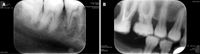
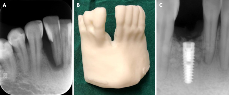
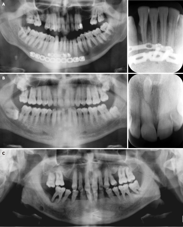
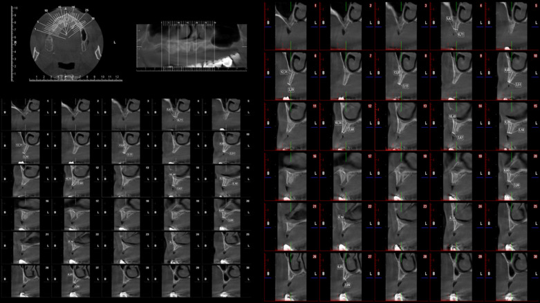
Oral and maxillofacial diagnostic imaging is of paramount importance in dental clinical diagnosis, treatment planning, and follow-up procedures. Periapical radiographic examination and numerous panoramic systems are used in routine clinical dental practice. Cone beam CT is widely used and currently the method of choice in oral and maxillofacial implantology, endodontics, maxillofacial surgery, periodontics, degenerative temporomandibular joint disease, orthodontics, airway studies, sleep disorders, and forensic dentistry. Another innovative laboratory research tool that offers three-dimensional (3D) detailed high-resolution images of teeth and neighboring structures with submicrometric accuracy is microcomputed tomography. Ultra-high radiation doses, long scanning times, and high costs preclude its routine clinical use. In response to the high demand for a technique that could provide real-time images using a cost-effective, rapid, user-friendly, and portable technique without ionizing radiation, some authors proposed ultrasound imaging methods as an alternative to X-ray imaging techniques. Ultrasonography can be used in the dentomaxillofacial region for various diagnostic purposes such as salivary gland and superficial tissue examination. Recently, dedicated dental magnetic resonance imaging with appropriate software, hardware, sequences, and field of view tailored to fit dentomaxillofacial anatomy was introduced. Lately, 3D printing technologies and their application in dentistry has attracted attention. During 3D printing a given material is added in successive layers to create a 3D object. The application of this technology has the potential to decrease operation time and minimize operator bias and the possibility of procedural errors. Another hot topic regarding dentomaxillofacial radiology is artificial intelligence, which is a field related to computer science dedicated to developing systems or machines that can perform tasks traditionally associated with human intelligence. It is obvious that further investigation and research in the field of dentomaxillofacial radiology will make great contributions to diagnostic imaging for various dental specialties.
口腔颌面诊断影像学在牙科临床诊断、治疗计划制定及随访程序中至关重要。根尖片检查及众多全景系统用于常规牙科临床实践。锥形束CT被广泛应用,目前是口腔颌面种植学、牙髓病学、颌面外科、牙周病学、颞下颌关节退行性疾病、正畸学、气道研究、睡眠障碍及法医牙科学的首选方法。另一种创新的实验室研究工具是显微计算机断层扫描,它能以亚微米精度提供牙齿及邻近结构的三维(3D)详细高分辨率图像。超高辐射剂量、长扫描时间及高成本使其无法用于常规临床。为满足对一种能使用经济高效、快速、用户友好且便携的无电离辐射技术提供实时图像的技术的高需求,一些作者提出超声成像方法作为X射线成像技术的替代方案。超声检查可用于牙颌面区域的各种诊断目的,如唾液腺和浅表组织检查。最近,引入了配备适合牙颌面解剖结构的适当软件、硬件、序列和视野的专用牙科磁共振成像。近来,3D打印技术及其在牙科中的应用引起了关注。在3D打印过程中,给定材料逐层添加以创建3D物体。该技术的应用有可能减少手术时间,并将操作员偏差和程序错误的可能性降至最低。牙颌面放射学的另一个热门话题是人工智能,它是计算机科学的一个领域,致力于开发能够执行传统上与人类智能相关任务的系统或机器。显然,牙颌面放射学领域的进一步研究将为各种牙科专业的诊断成像做出巨大贡献。