• 文献检索
  • 文档翻译
  • 深度研究
  • 学术资讯
  • Suppr Zotero 插件Zotero 插件
  • 邀请有礼
  • 套餐&价格
  • 历史记录
应用&插件
Suppr Zotero 插件Zotero 插件浏览器插件Mac 客户端Windows 客户端微信小程序
定价
高级版会员购买积分包购买API积分包
服务
文献检索文档翻译深度研究API 文档MCP 服务
关于我们
关于 Suppr公司介绍联系我们用户协议隐私条款
关注我们

Suppr 超能文献

核心技术专利:CN118964589B侵权必究
粤ICP备2023148730 号-1Suppr @ 2026

文献检索

告别复杂PubMed语法,用中文像聊天一样搜索,搜遍4000万医学文献。AI智能推荐,让科研检索更轻松。

立即免费搜索

文件翻译

保留排版,准确专业,支持PDF/Word/PPT等文件格式,支持 12+语言互译。

免费翻译文档

深度研究

AI帮你快速写综述,25分钟生成高质量综述,智能提取关键信息,辅助科研写作。

立即免费体验

用于组装组织初级纤毛的远端附属器的分级途径。

A hierarchical pathway for assembly of the distal appendages that organize primary cilia.

作者信息

Kanie Tomoharu, Liu Beibei, Love Julia F, Fisher Saxton D, Gustavsson Anna-Karin, Jackson Peter K

机构信息

Baxter Laboratory, Department of Microbiology & Immunology and Department of Pathology, Stanford University, Stanford, United States.

Department of Cell Biology, University of Oklahoma Health Sciences Center, Oklahoma City, United States.

出版信息

Elife. 2025 Jan 30;14:e85999. doi: 10.7554/eLife.85999.

DOI:10.7554/eLife.85999
PMID:39882846
原文链接:https://pmc.ncbi.nlm.nih.gov/articles/PMC11984956/
Abstract

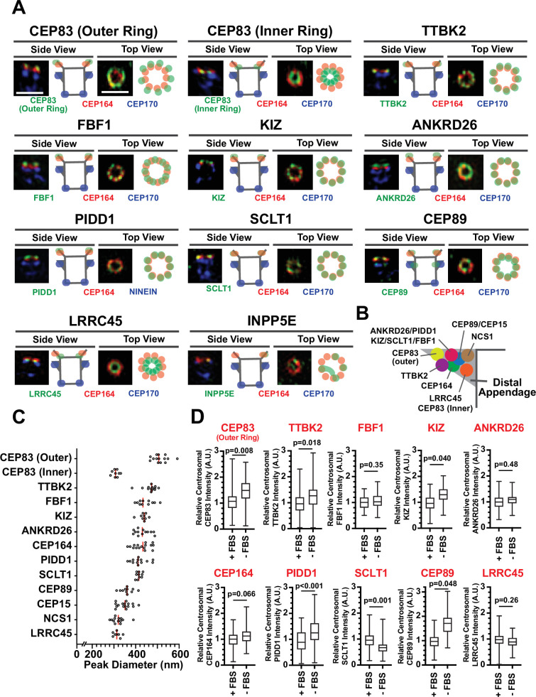
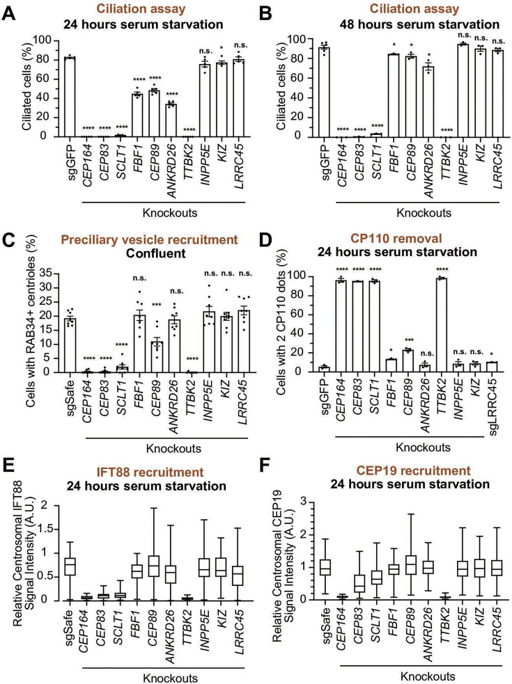
Distal appendages are ninefold symmetric blade-like structures attached to the distal end of the mother centriole. These structures are critical for the formation of the primary cilium, by regulating at least four critical steps: preciliary vesicle recruitment, recruitment and initiation of intraflagellar transport (IFT), and removal of CP110. While specific proteins that localize to the distal appendages have been identified, how exactly each protein functions to achieve the multiple roles of the distal appendages is poorly understood. Here, we comprehensively analyze known and newly discovered distal appendage proteins (CEP83, SCLT1, CEP164, TTBK2, FBF1, CEP89, KIZ, ANKRD26, PIDD1, LRRC45, NCS1, CEP15) for their precise localization, order of recruitment, and their roles in each step of cilia formation. Using CRISPR-Cas9 knockouts, we show that the order of the recruitment of the distal appendage proteins is highly interconnected and a more complex hierarchy. Our analysis highlights two protein modules, CEP83-SCLT1 and CEP164-TTBK2, as critical for structural assembly of distal appendages. Functional assays revealed that CEP89 selectively functions in the RAB34 vesicle recruitment, while deletion of the integral components, CEP83-SCLT1-CEP164-TTBK2, severely compromised all four steps of cilium formation. Collectively, our analyses provide a more comprehensive view of the organization and the function of the distal appendage, paving the way for molecular understanding of ciliary assembly.

摘要

远端附属物是附着于母中心粒远端的九重对称叶片状结构。这些结构对于初级纤毛的形成至关重要,通过调节至少四个关键步骤:纤毛前囊泡募集、鞭毛内运输(IFT)的募集和起始,以及CP110的去除。虽然已经鉴定出定位于远端附属物的特定蛋白质,但每种蛋白质究竟如何发挥作用以实现远端附属物的多种功能却知之甚少。在这里,我们全面分析了已知的和新发现的远端附属物蛋白(CEP83、SCLT1、CEP164、TTBK2、FBF1、CEP89、KIZ、ANKRD26、PIDD1、LRRC45、NCS1、CEP15)的精确定位、募集顺序及其在纤毛形成各步骤中的作用。使用CRISPR-Cas9基因敲除技术,我们表明远端附属物蛋白的募集顺序高度相互关联且层次更为复杂。我们的分析突出了两个蛋白质模块,即CEP83-SCLT1和CEP164-TTBK2,对远端附属物的结构组装至关重要。功能分析显示,CEP89在RAB34囊泡募集中具有选择性功能,而缺失整合成分CEP83-SCLT1-CEP164-TTBK2会严重损害纤毛形成的所有四个步骤。总的来说,我们的分析为远端附属物的组织和功能提供了更全面的视角,为纤毛组装的分子理解铺平了道路。

https://cdn.ncbi.nlm.nih.gov/pmc/blobs/4a14/11984956/650680774d3d/elife-85999-sa2-fig3.jpg
https://cdn.ncbi.nlm.nih.gov/pmc/blobs/4a14/11984956/0c23d1da5b58/elife-85999-fig1.jpg
https://cdn.ncbi.nlm.nih.gov/pmc/blobs/4a14/11984956/90b1d635a48b/elife-85999-fig1-figsupp1.jpg
https://cdn.ncbi.nlm.nih.gov/pmc/blobs/4a14/11984956/284b211c658d/elife-85999-fig1-figsupp2.jpg
https://cdn.ncbi.nlm.nih.gov/pmc/blobs/4a14/11984956/3358b9533668/elife-85999-fig1-figsupp3.jpg
https://cdn.ncbi.nlm.nih.gov/pmc/blobs/4a14/11984956/370423606cf6/elife-85999-fig1-figsupp4.jpg
https://cdn.ncbi.nlm.nih.gov/pmc/blobs/4a14/11984956/33c2e5050402/elife-85999-fig1-figsupp5.jpg
https://cdn.ncbi.nlm.nih.gov/pmc/blobs/4a14/11984956/bb7ef3e69836/elife-85999-fig2.jpg
https://cdn.ncbi.nlm.nih.gov/pmc/blobs/4a14/11984956/d71f15039dd2/elife-85999-fig2-figsupp1.jpg
https://cdn.ncbi.nlm.nih.gov/pmc/blobs/4a14/11984956/d8a2ac361ace/elife-85999-fig2-figsupp2.jpg
https://cdn.ncbi.nlm.nih.gov/pmc/blobs/4a14/11984956/507df9b93766/elife-85999-fig3.jpg
https://cdn.ncbi.nlm.nih.gov/pmc/blobs/4a14/11984956/091032576e84/elife-85999-fig3-figsupp1.jpg
https://cdn.ncbi.nlm.nih.gov/pmc/blobs/4a14/11984956/8101ea9bcf02/elife-85999-fig3-figsupp2.jpg
https://cdn.ncbi.nlm.nih.gov/pmc/blobs/4a14/11984956/3158b65b3f3e/elife-85999-fig3-figsupp3.jpg
https://cdn.ncbi.nlm.nih.gov/pmc/blobs/4a14/11984956/f572eb867430/elife-85999-fig3-figsupp4.jpg
https://cdn.ncbi.nlm.nih.gov/pmc/blobs/4a14/11984956/ce7288c123dd/elife-85999-fig3-figsupp5.jpg
https://cdn.ncbi.nlm.nih.gov/pmc/blobs/4a14/11984956/458ac929a291/elife-85999-fig3-figsupp6.jpg
https://cdn.ncbi.nlm.nih.gov/pmc/blobs/4a14/11984956/8eb8c1470e24/elife-85999-fig3-figsupp7.jpg
https://cdn.ncbi.nlm.nih.gov/pmc/blobs/4a14/11984956/6d0c4786c569/elife-85999-fig4.jpg
https://cdn.ncbi.nlm.nih.gov/pmc/blobs/4a14/11984956/fbf9965437ff/elife-85999-fig4-figsupp1.jpg
https://cdn.ncbi.nlm.nih.gov/pmc/blobs/4a14/11984956/9d1ca6870fb7/elife-85999-fig4-figsupp2.jpg
https://cdn.ncbi.nlm.nih.gov/pmc/blobs/4a14/11984956/f984ebd1be08/elife-85999-fig5.jpg
https://cdn.ncbi.nlm.nih.gov/pmc/blobs/4a14/11984956/7a188d47dfdf/elife-85999-fig5-figsupp1.jpg
https://cdn.ncbi.nlm.nih.gov/pmc/blobs/4a14/11984956/1bf463b06f73/elife-85999-fig6.jpg
https://cdn.ncbi.nlm.nih.gov/pmc/blobs/4a14/11984956/137de019986a/elife-85999-sa2-fig1.jpg
https://cdn.ncbi.nlm.nih.gov/pmc/blobs/4a14/11984956/60a0b0d26ac6/elife-85999-sa2-fig2.jpg
https://cdn.ncbi.nlm.nih.gov/pmc/blobs/4a14/11984956/650680774d3d/elife-85999-sa2-fig3.jpg

相似文献

1
A hierarchical pathway for assembly of the distal appendages that organize primary cilia.用于组装组织初级纤毛的远端附属器的分级途径。
Elife. 2025 Jan 30;14:e85999. doi: 10.7554/eLife.85999.
2
A hierarchical pathway for assembly of the distal appendages that organize primary cilia.用于组装组织初级纤毛的远端附属器的分级途径。
bioRxiv. 2023 Jan 10:2023.01.06.522944. doi: 10.1101/2023.01.06.522944.
3
Centriole distal appendages promote membrane docking, leading to cilia initiation.中心粒远端附属物促进膜对接,从而引发纤毛起始。
Genes Dev. 2013 Jan 15;27(2):163-8. doi: 10.1101/gad.207043.112.
4
Myristoylated Neuronal Calcium Sensor-1 captures the preciliary vesicle at distal appendages.肉豆蔻酰化的神经元钙传感器-1在远端附属物处捕获纤毛前小泡。
Elife. 2025 Jan 30;14:e85998. doi: 10.7554/eLife.85998.
5
C2cd3 is critical for centriolar distal appendage assembly and ciliary vesicle docking in mammals.C2cd3 对于中心粒远端附属物的组装和纤毛囊泡的对接在哺乳动物中是至关重要的。
Proc Natl Acad Sci U S A. 2014 Feb 11;111(6):2164-9. doi: 10.1073/pnas.1318737111. Epub 2014 Jan 27.
6
LRRC45 contributes to early steps of axoneme extension.LRRC45 有助于轴丝延伸的早期步骤。
J Cell Sci. 2018 Sep 20;131(18):jcs223594. doi: 10.1242/jcs.223594.
7
Super-resolution architecture of mammalian centriole distal appendages reveals distinct blade and matrix functional components.哺乳动物中心粒远端附属物的超分辨率结构揭示了不同的叶片和基质功能组件。
Nat Commun. 2018 May 22;9(1):2023. doi: 10.1038/s41467-018-04469-1.
8
Cep164 triggers ciliogenesis by recruiting Tau tubulin kinase 2 to the mother centriole.CEP164 通过将 Tau 微管激酶 2 招募到母中心粒来触发纤毛发生。
Proc Natl Acad Sci U S A. 2014 Jul 15;111(28):E2841-50. doi: 10.1073/pnas.1401777111. Epub 2014 Jun 30.
9
Distinct roles of centriole distal appendage proteins in ciliary assembly and disassembly.中心粒远端附属蛋白在纤毛组装和解聚中的不同作用。
Cell Commun Signal. 2024 Dec 18;22(1):607. doi: 10.1186/s12964-024-01962-7.
10
Phosphorylation of CEP83 by TTBK2 is necessary for cilia initiation.CEP83 由 TTBK2 磷酸化对于纤毛起始是必要的。
J Cell Biol. 2019 Oct 7;218(10):3489-3505. doi: 10.1083/jcb.201811142. Epub 2019 Aug 27.

引用本文的文献

1
Characterization of membrane structures regulating primary ciliogenesis by quantitative isotropic ultrastructure imaging.通过定量各向同性超微结构成像对调节初级纤毛发生的膜结构进行表征。
bioRxiv. 2025 Aug 21:2025.08.20.670930. doi: 10.1101/2025.08.20.670930.
2
Single-objective lattice light sheet microscopy with microfluidics for single-molecule super-resolution imaging of mammalian cells.用于哺乳动物细胞单分子超分辨率成像的带微流控的单目标晶格光片显微镜
bioRxiv. 2025 Jul 23:2025.07.18.665606. doi: 10.1101/2025.07.18.665606.
3
Novel reporter systems to detect cold and osmotic stress responses.

本文引用的文献

1
Myristoylated Neuronal Calcium Sensor-1 captures the preciliary vesicle at distal appendages.肉豆蔻酰化的神经元钙传感器-1在远端附属物处捕获纤毛前小泡。
Elife. 2025 Jan 30;14:e85998. doi: 10.7554/eLife.85998.
2
Identification of a third myosin-5a-melanophilin interaction that mediates the association of myosin-5a with melanosomes.鉴定介导肌球蛋白-5a 与黑素体结合的第三个肌球蛋白-5a-黑素磷蛋白相互作用。
Elife. 2024 Jun 20;13:RP93662. doi: 10.7554/eLife.93662.
3
Highly accurate protein structure prediction with AlphaFold.利用 AlphaFold 进行高精度蛋白质结构预测。
用于检测冷应激和渗透应激反应的新型报告系统。
Biol Methods Protoc. 2025 Jun 14;10(1):bpaf048. doi: 10.1093/biomethods/bpaf048. eCollection 2025.
4
Cilia at the crossroad: convergence of regulatory mechanisms to govern cilia dynamics during cell signaling and the cell cycle.纤毛处于十字路口:细胞信号传导和细胞周期中调控纤毛动态的调节机制的汇聚
Cell Biosci. 2025 Jun 7;15(1):81. doi: 10.1186/s13578-025-01403-z.
5
Synchronized temporal-spatial analysis via microscopy and phosphoproteomics (STAMP) of quiescence.通过显微镜和磷酸化蛋白质组学对静止状态进行同步时空分析(STAMP)。
Sci Adv. 2025 Apr 25;11(17):eadt9712. doi: 10.1126/sciadv.adt9712.
6
Tau-tubulin kinase 2 restrains microtubule-depolymerizer KIF2A to support primary cilia growth.微管蛋白激酶2抑制微管解聚酶KIF2A以支持初级纤毛生长。
Cell Commun Signal. 2025 Feb 10;23(1):73. doi: 10.1186/s12964-025-02072-8.
7
Myristoylated Neuronal Calcium Sensor-1 captures the preciliary vesicle at distal appendages.肉豆蔻酰化的神经元钙传感器-1在远端附属物处捕获纤毛前小泡。
Elife. 2025 Jan 30;14:e85998. doi: 10.7554/eLife.85998.
8
Biallelic Variants in LRRC45 Impair Ciliogenesis and Cause a Severe Neurological Disorder.LRRC45基因的双等位基因变异损害纤毛生成并导致严重的神经疾病。
Clin Genet. 2025 Mar;107(3):311-322. doi: 10.1111/cge.14663. Epub 2024 Dec 5.
9
Disruption of distal appendage protein CEP164 causes skeletal malformation in mice.远端附属蛋白CEP164的破坏导致小鼠骨骼畸形。
Biochem Biophys Res Commun. 2024 Dec 31;741:151063. doi: 10.1016/j.bbrc.2024.151063. Epub 2024 Nov 26.
10
Long axial-range double-helix point spread functions for 3D volumetric super-resolution imaging.用于三维体积超分辨率成像的长轴向范围双螺旋点扩散函数
bioRxiv. 2024 Aug 1:2024.07.31.605907. doi: 10.1101/2024.07.31.605907.
Nature. 2021 Aug;596(7873):583-589. doi: 10.1038/s41586-021-03819-2. Epub 2021 Jul 15.
4
Seeing beyond the limit: A guide to choosing the right super-resolution microscopy technique.超越极限:选择合适的超分辨率显微镜技术指南。
J Biol Chem. 2021 Jul;297(1):100791. doi: 10.1016/j.jbc.2021.100791. Epub 2021 May 18.
5
Rab34 GTPase mediates ciliary membrane formation in the intracellular ciliogenesis pathway.Rab34 GTPase 介导细胞内纤毛发生途径中的纤毛膜形成。
Curr Biol. 2021 Jul 12;31(13):2895-2905.e7. doi: 10.1016/j.cub.2021.04.075. Epub 2021 May 13.
6
Rab34 is necessary for early stages of intracellular ciliogenesis.Rab34 对于细胞内纤毛发生的早期阶段是必需的。
Curr Biol. 2021 Jul 12;31(13):2887-2894.e4. doi: 10.1016/j.cub.2021.04.018. Epub 2021 May 13.
7
ANKRD26 recruits PIDD1 to centriolar distal appendages to activate the PIDDosome following centrosome amplification.ANKRD26 将 PIDD1 招募到中心粒远端附属物,以在中心体扩增后激活 PIDDosome。
EMBO J. 2021 Feb 15;40(4):e105106. doi: 10.15252/embj.2020105106. Epub 2020 Dec 22.
8
Centriolar distal appendages activate the centrosome-PIDDosome-p53 signalling axis via ANKRD26.中心粒远端附属物通过 ANKRD26 激活中心体-PIDDosome-p53 信号轴。
EMBO J. 2021 Feb 15;40(4):e104844. doi: 10.15252/embj.2020104844. Epub 2020 Dec 22.
9
SMAP: a modular super-resolution microscopy analysis platform for SMLM data.SMAP:用于单分子定位显微镜(SMLM)数据的模块化超分辨率显微镜分析平台。
Nat Methods. 2020 Sep;17(9):870-872. doi: 10.1038/s41592-020-0938-1.
10
A comprehensive analysis of Rab GTPases reveals a role for Rab34 in serum starvation-induced primary ciliogenesis.全面分析 Rab GTPases 揭示了 Rab34 在血清饥饿诱导的初级纤毛生成中的作用。
J Biol Chem. 2020 Sep 4;295(36):12674-12685. doi: 10.1074/jbc.RA119.012233. Epub 2020 Jul 15.