Cacciaguerra Laura, Storelli Loredana, Pagani Elisabetta, Preziosa Paolo, Mesaros Sharlota, Martinelli Vittorio, Moiola Lucia, Radaelli Marta, Ivanovic Jovana, Tamas Olivera, Drulovic Jelena, Filippi Massimo, Rocca Maria A
Neuroimaging Research Unit, Division of Neuroscience, IRCCS San Raffaele Scientific Institute, Milan, Italy; Vita-Salute San Raffaele University, Milan, Italy.
Neuroimaging Research Unit, Division of Neuroscience, IRCCS San Raffaele Scientific Institute, Milan, Italy.
Mult Scler. 2025 Feb;31(2):140-158. doi: 10.1177/13524585241307154. Epub 2024 Dec 31.
The understanding of disease pathophysiology is pivotal for tailored treatments. The spatial distribution of brain damage relies on the regional antigen expression and the local balance of susceptibility and protective elements.
As proof-of-concept, we investigated the spatial association between brain damage and gene expression in aquaporin-4-IgG-positive neuromyelitis optica spectrum disorder (AQP4 + NMOSD).
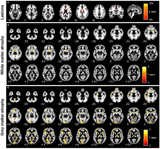
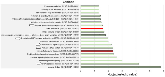
In this multicenter cross-sectional study, 90 AQP4 + NMOSD patients and 94 age-matched healthy controls underwent a brain magnetic resonance imaging (MRI). We used T2-hyperintense lesion probability maps and white/gray matter atrophy as proxies of inflammation and neurodegeneration. The association with the expression of 266 candidate genes was obtained with the Multimodal Environment for Neuroimaging and Genomic Analysis platform. A functional-enrichment analysis investigated overrepresented biological processes.
In AQP4 + NMOSD, T2-hyperintense lesions were mainly periventricular; atrophy mostly involved the visual pathway. The expression of AQP4 and complement (C4a and C5) was associated with both inflammation and neurodegeneration. Complement activation and regulation/uptake of the insulin-like growth factor were the most relevant enriched pathways. Nonspecific pathways related to DNA synthesis and repair were associated with brain atrophy.
Quantitative MRI and gene expression atlas identified the key elements of AQP4 + NMOSD pathophysiology. This analysis could help in understanding the pathophysiology of antibody-mediated autoimmune disorders.
对疾病病理生理学的理解对于个体化治疗至关重要。脑损伤的空间分布依赖于区域抗原表达以及易感性和保护因素的局部平衡。
作为概念验证,我们研究了水通道蛋白4-IgG阳性视神经脊髓炎谱系障碍(AQP4+NMOSD)中脑损伤与基因表达之间的空间关联。
在这项多中心横断面研究中,90例AQP4+NMOSD患者和94例年龄匹配的健康对照者接受了脑磁共振成像(MRI)检查。我们使用T2高信号病变概率图以及白质/灰质萎缩作为炎症和神经退行性变的替代指标。通过神经影像学和基因组分析多模态环境平台获得与266个候选基因表达的关联。功能富集分析研究了过度表达的生物学过程。
在AQP4+NMOSD中,T2高信号病变主要位于脑室周围;萎缩主要累及视觉通路。AQP4和补体(C4a和C5)的表达与炎症和神经退行性变均相关。补体激活以及胰岛素样生长因子的调节/摄取是最相关的富集通路。与DNA合成和修复相关的非特异性通路与脑萎缩有关。
定量MRI和基因表达图谱确定了AQP4+NMOSD病理生理学的关键要素。该分析有助于理解抗体介导的自身免疫性疾病的病理生理学。