• 文献检索
  • 文档翻译
  • 深度研究
  • 学术资讯
  • Suppr Zotero 插件Zotero 插件
  • 邀请有礼
  • 套餐&价格
  • 历史记录
应用&插件
Suppr Zotero 插件Zotero 插件浏览器插件Mac 客户端Windows 客户端微信小程序
定价
高级版会员购买积分包购买API积分包
服务
文献检索文档翻译深度研究API 文档MCP 服务
关于我们
关于 Suppr公司介绍联系我们用户协议隐私条款
关注我们

Suppr 超能文献

核心技术专利:CN118964589B侵权必究
粤ICP备2023148730 号-1Suppr @ 2026

文献检索

告别复杂PubMed语法,用中文像聊天一样搜索,搜遍4000万医学文献。AI智能推荐,让科研检索更轻松。

立即免费搜索

文件翻译

保留排版,准确专业,支持PDF/Word/PPT等文件格式,支持 12+语言互译。

免费翻译文档

深度研究

AI帮你快速写综述,25分钟生成高质量综述,智能提取关键信息,辅助科研写作。

立即免费体验

肝细胞中RelA信号受损与脂质代谢失调:代谢功能障碍相关脂肪性肝病进展的驱动因素

Impaired RelA signaling and lipid metabolism dysregulation in hepatocytes: driving forces in the progression of metabolic dysfunction-associated steatotic liver disease.

作者信息

He Yihuai, Jiang Jinlian, Ou Lili, Chen Yunfen, Abudukeremu Aikedaimu, Chen Guimei, Zhong Weiwei, Jiang Zhigang, Nuermaimaiti Nuerbiye, Guan Yaqun

机构信息

State Key Laboratory of Pathogenesis, Prevention and Treatment of High Incidence Diseases in Central Asia, Xinjiang Key Laboratory of Molecular Biology for Endemic Diseases, Department of Pathology, School of Basic Medical Sciences, Xinjiang Medical University, Urumqi, Xinjiang, China.

Department of Infectious Diseases, Affiliated Hospital of Zunyi Medical University, Zunyi, Guizhou, China.

出版信息

Cell Death Discov. 2025 Feb 5;11(1):49. doi: 10.1038/s41420-025-02312-3.

DOI:10.1038/s41420-025-02312-3
PMID:39910053
原文链接:https://pmc.ncbi.nlm.nih.gov/articles/PMC11799324/
Abstract

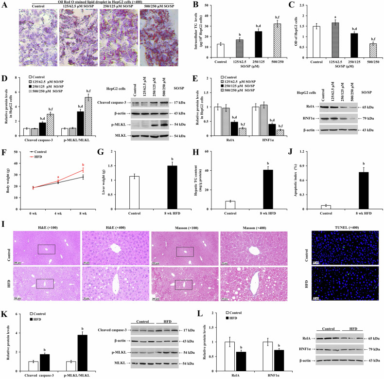
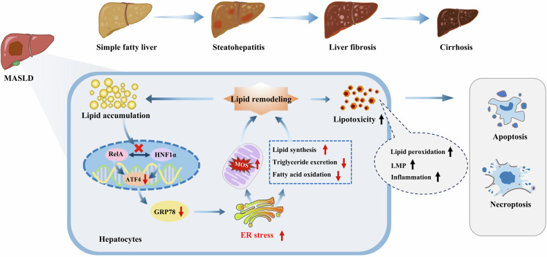
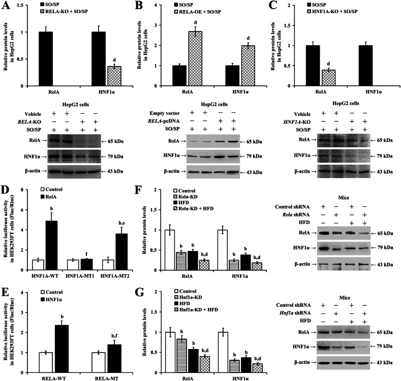
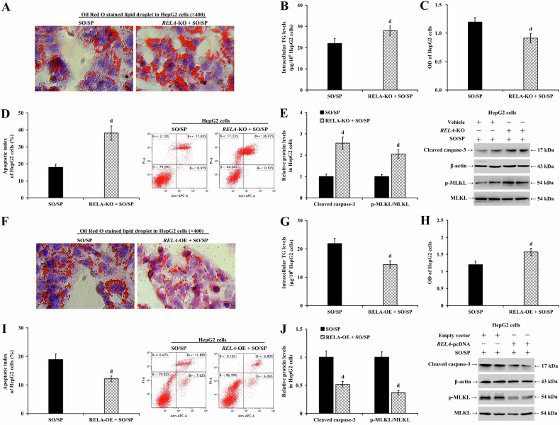
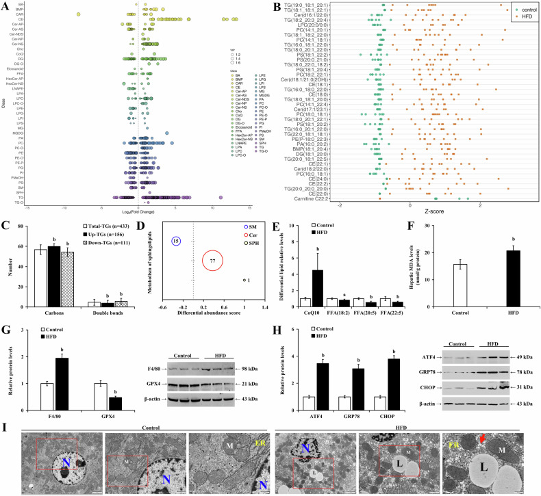
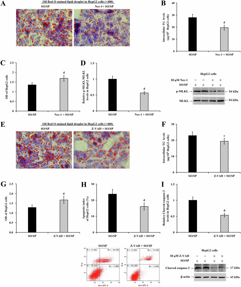
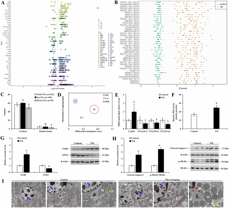
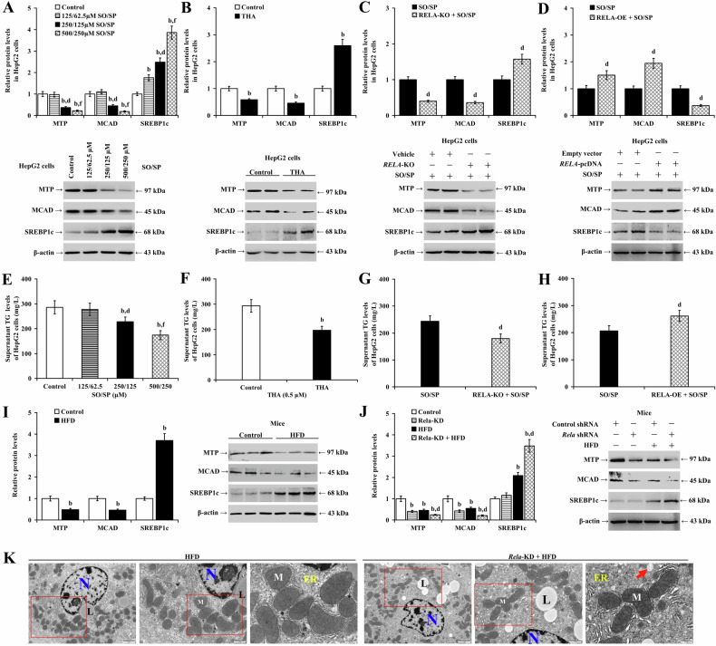
RelA, also known as nuclear factor kappa B p65, plays a crucial role in the pathogenesis of various liver diseases. However, the specific role of RelA in hepatocytes during the progression of metabolic dysfunction-associated steatotic liver disease (MASLD) is not well understood. This study explored the relationship between impaired RelA signaling and lipid metabolism disorders in hepatocytes, and how they synergistically contribute to the advancement of MASLD. We assessed the changes, regulatory relationships, and impacts of RelA signaling and lipid metabolism remodeling on disease progression both in vitro and in vivo. During MASLD, there was a decrease in the expression of RelA and hepatocyte nuclear factor 1 alpha (HNF1α), with both factors showing mutual enhancement of each other's expression under normal conditions. This synergistic effect was absent during hepatocyte steatosis. RelA or HNF1α depletion in hepatocytes intensified MASLD symptoms, whereas overexpression of RELA or treatment with necrostatin-1 (a necroptosis inhibitor) or Z-VAD (a caspase inhibitor) significantly mitigated these effects. Mechanistically, during hepatic steatosis, altered lipid profiles exhibited lipotoxicity, inducing hepatocyte apoptosis and necroptosis, whereas endoplasmic reticulum (ER) stress triggered lipid remodeling processes similar to those observed in MASLD. RelA signaling upregulated the expression of activating transcription factor 4 and glucose-regulated protein 78, thereby alleviating ER stress. Impaired RelA signaling remodeled the ER stress response and lipid metabolism, and enhanced lipid accumulation and lipid toxicity. In conclusion, impaired RelA signaling and disrupted lipid metabolism form a detrimental feedback loop in hepatocytes that promotes MASLD progression. Lipid accumulation suppresses RelA signaling, remodeling the ER stress response and exacerbating lipid metabolism disorder, ultimately leading to hepatocyte apoptosis and necroptosis.

摘要

RelA,也被称为核因子κB p65,在各种肝脏疾病的发病机制中起着关键作用。然而,RelA在代谢功能障碍相关脂肪性肝病(MASLD)进展过程中在肝细胞中的具体作用尚不清楚。本研究探讨了RelA信号受损与肝细胞脂质代谢紊乱之间的关系,以及它们如何协同促进MASLD的进展。我们在体外和体内评估了RelA信号和脂质代谢重塑对疾病进展的变化、调控关系及影响。在MASLD期间,RelA和肝细胞核因子1α(HNF1α)的表达下降,而在正常条件下这两个因子显示出相互增强彼此的表达。在肝细胞脂肪变性期间这种协同效应不存在。肝细胞中RelA或HNF1α的缺失加剧了MASLD症状,而RELA的过表达或用坏死性凋亡抑制剂坏死抑素-1或半胱天冬酶抑制剂Z-VAD处理则显著减轻了这些影响。机制上,在肝脂肪变性期间,改变的脂质谱表现出脂毒性,诱导肝细胞凋亡和坏死性凋亡,而内质网(ER)应激触发了类似于MASLD中观察到的脂质重塑过程。RelA信号上调激活转录因子4和葡萄糖调节蛋白78的表达,从而减轻ER应激。RelA信号受损重塑了ER应激反应和脂质代谢,并增强了脂质积累和脂质毒性。总之,RelA信号受损和脂质代谢紊乱在肝细胞中形成了一个有害的反馈回路,促进了MASLD的进展。脂质积累抑制RelA信号,重塑ER应激反应并加剧脂质代谢紊乱,最终导致肝细胞凋亡和坏死性凋亡。

https://cdn.ncbi.nlm.nih.gov/pmc/blobs/1f1f/11799324/23c2fa27ab9b/41420_2025_2312_Fig11_HTML.jpg
https://cdn.ncbi.nlm.nih.gov/pmc/blobs/1f1f/11799324/d00443db88a4/41420_2025_2312_Fig1_HTML.jpg
https://cdn.ncbi.nlm.nih.gov/pmc/blobs/1f1f/11799324/e2c6bd87ef83/41420_2025_2312_Fig2_HTML.jpg
https://cdn.ncbi.nlm.nih.gov/pmc/blobs/1f1f/11799324/a02d38156a92/41420_2025_2312_Fig3_HTML.jpg
https://cdn.ncbi.nlm.nih.gov/pmc/blobs/1f1f/11799324/fc4858f5b088/41420_2025_2312_Fig4_HTML.jpg
https://cdn.ncbi.nlm.nih.gov/pmc/blobs/1f1f/11799324/338d4aac1650/41420_2025_2312_Fig5_HTML.jpg
https://cdn.ncbi.nlm.nih.gov/pmc/blobs/1f1f/11799324/178c40c8c3ee/41420_2025_2312_Fig6_HTML.jpg
https://cdn.ncbi.nlm.nih.gov/pmc/blobs/1f1f/11799324/aff0b9f9cf96/41420_2025_2312_Fig7_HTML.jpg
https://cdn.ncbi.nlm.nih.gov/pmc/blobs/1f1f/11799324/49911c868c61/41420_2025_2312_Fig8_HTML.jpg
https://cdn.ncbi.nlm.nih.gov/pmc/blobs/1f1f/11799324/7ab2b1b89d02/41420_2025_2312_Fig9_HTML.jpg
https://cdn.ncbi.nlm.nih.gov/pmc/blobs/1f1f/11799324/dd50dfe082eb/41420_2025_2312_Fig10_HTML.jpg
https://cdn.ncbi.nlm.nih.gov/pmc/blobs/1f1f/11799324/23c2fa27ab9b/41420_2025_2312_Fig11_HTML.jpg

相似文献

1
Impaired RelA signaling and lipid metabolism dysregulation in hepatocytes: driving forces in the progression of metabolic dysfunction-associated steatotic liver disease.肝细胞中RelA信号受损与脂质代谢失调:代谢功能障碍相关脂肪性肝病进展的驱动因素
Cell Death Discov. 2025 Feb 5;11(1):49. doi: 10.1038/s41420-025-02312-3.
2
polysaccharides alleviate metabolic dysfunction-associated steatotic liver disease through enhancing hepatocyte RelA/ HNF1α signaling.多糖通过增强肝细胞RelA/HNF1α信号传导来缓解代谢功能障碍相关的脂肪性肝病。
World J Gastroenterol. 2025 Jan 28;31(4):93179. doi: 10.3748/wjg.v31.i4.93179.
3
Feedback loop between hepatocyte nuclear factor 1α and endoplasmic reticulum stress mitigates liver injury by downregulating hepatocyte apoptosis.肝细胞核因子 1α 与内质网应激之间的反馈环通过下调肝细胞凋亡减轻肝损伤。
Sci Rep. 2022 Jul 8;12(1):11602. doi: 10.1038/s41598-022-15846-8.
4
Cell Homeostasis or Cell Death-The Balancing Act Between Autophagy and Apoptosis Caused by Steatosis-Induced Endoplasmic Reticulum (ER) Stress.细胞稳态或细胞死亡——脂肪变性诱导的内质网(ER)应激引起的自噬与凋亡之间的平衡行为
Cells. 2025 Mar 18;14(6):449. doi: 10.3390/cells14060449.
5
Caffeic acid ameliorates metabolic dysfunction-associated steatotic liver disease via alleviating oxidative damage and lipid accumulation in hepatocytes through activating Nrf2 via targeting Keap1.咖啡酸通过靶向 Keap1 激活 Nrf2 减轻肝细胞氧化损伤和脂质堆积来改善代谢相关脂肪性肝病。
Free Radic Biol Med. 2024 Nov 1;224:352-365. doi: 10.1016/j.freeradbiomed.2024.08.038. Epub 2024 Aug 28.
6
A read-through circular RNA RCRIN inhibits metabolic dysfunction-associated steatotic liver disease.一种通读环状RNA RCRIN可抑制代谢功能障碍相关脂肪性肝病。
J Hepatol. 2025 Jun;82(6):1068-1079. doi: 10.1016/j.jhep.2024.11.052. Epub 2024 Dec 10.
7
YBX1/CD36 positive feedback loop-mediated lipid accumulation drives metabolic dysfunction-associated steatotic liver disease.YBX1/CD36 正反馈回路介导的脂质积累驱动代谢功能障碍相关脂肪性肝病。
Int J Biol Sci. 2025 Feb 18;21(5):2118-2134. doi: 10.7150/ijbs.105798. eCollection 2025.
8
Hepatic miR-149-5p upregulation fosters steatosis, inflammation and fibrosis development in mice and in human liver organoids.肝脏中miR-149-5p的上调促进小鼠和人肝脏类器官中的脂肪变性、炎症和纤维化发展。
JHEP Rep. 2024 Jun 4;6(9):101126. doi: 10.1016/j.jhepr.2024.101126. eCollection 2024 Sep.
9
Hepatocyte-specific Epidermal Growth Factor Receptor Deletion Promotes Fibrosis but has no Effect on Steatosis in Fast-food Diet Model of Metabolic Dysfunction-associated Steatotic Liver Disease.肝细胞特异性表皮生长因子受体缺失促进肝纤维化,但对代谢相关脂肪性肝病的快餐饮食模型中的脂肪变性没有影响。
Cell Mol Gastroenterol Hepatol. 2024;18(4):101380. doi: 10.1016/j.jcmgh.2024.101380. Epub 2024 Jul 20.
10
Natural linoleic acid from marine fungus Eutypella sp. F0219 blocks KEAP1/NRF2 interaction and ameliorates MASLD by targeting FABP4.海洋真菌 Eutypella sp. F0219 中的天然亚油酸通过靶向 FABP4 阻断 KEAP1/NRF2 相互作用并改善 MASLD。
Free Radic Biol Med. 2024 Nov 1;224:630-643. doi: 10.1016/j.freeradbiomed.2024.09.019. Epub 2024 Sep 17.

引用本文的文献

1
Specnuezhenide mitigates tunicamycin-induced liver injury in mice via ER stress modulation and metabolic reprogramming.蛇床子素通过内质网应激调节和代谢重编程减轻衣霉素诱导的小鼠肝损伤。
Mol Biol Rep. 2025 Aug 11;52(1):814. doi: 10.1007/s11033-025-10920-6.
2
Identification of palmitoylated biomarkers in non-alcoholic fatty liver disease via integrated bioinformatics analysis and machine learning.通过综合生物信息学分析和机器学习鉴定非酒精性脂肪性肝病中的棕榈酰化生物标志物
Sci Rep. 2025 Aug 1;15(1):28177. doi: 10.1038/s41598-025-13477-3.

本文引用的文献

1
Endoplasmic reticulum: Monitoring and maintaining protein and membrane homeostasis in the endoplasmic reticulum by the unfolded protein response.内质网:未折叠蛋白反应对内质网中蛋白质和膜稳态的监测和维持。
Int J Biochem Cell Biol. 2024 Jul;172:106598. doi: 10.1016/j.biocel.2024.106598. Epub 2024 May 18.
2
Activating transcription factor 4: a regulator of stress response in human cancers.激活转录因子4:人类癌症应激反应的调节因子
Front Cell Dev Biol. 2024 Mar 27;12:1370012. doi: 10.3389/fcell.2024.1370012. eCollection 2024.
3
Uridine diphosphate glucuronosyltransferase 1A1 prevents the progression of liver injury.
尿苷二磷酸葡萄糖醛酸基转移酶 1A1 可阻止肝损伤的进展。
World J Gastroenterol. 2024 Mar 7;30(9):1189-1212. doi: 10.3748/wjg.v30.i9.1189.
4
Acute endoplasmic reticulum stress-induced mitochondria respiratory chain damage: The role of activated calpains.急性内质网应激诱导的线粒体呼吸链损伤:活化钙蛋白酶的作用
FASEB J. 2024 Jan 31;38(2):e23404. doi: 10.1096/fj.202301158RR.
5
Nonalcoholic Fatty Liver Disease Gets Renamed as Metabolic Dysfunction-Associated Steatotic Liver Disease: Progress But With Challenges.非酒精性脂肪性肝病更名为代谢功能障碍相关脂肪性肝病:进展与挑战并存
Gastroenterology. 2024 Feb;166(2):229-234. doi: 10.1053/j.gastro.2023.11.007. Epub 2023 Nov 8.
6
Natural history of metabolic dysfunction-associated steatotic liver disease.代谢功能障碍相关脂肪性肝病的自然史。
Eur J Intern Med. 2024 Apr;122:3-10. doi: 10.1016/j.ejim.2023.11.005. Epub 2023 Nov 7.
7
Dominant-negative HNF1α mutant promotes liver steatosis and inflammation by regulating hepatic complement factor D.显性负性HNF1α突变体通过调节肝脏补体因子D促进肝脂肪变性和炎症。
iScience. 2023 Sep 22;26(10):108018. doi: 10.1016/j.isci.2023.108018. eCollection 2023 Oct 20.
8
Structural rather than catalytic role for mitochondrial respiratory chain supercomplexes.线粒体呼吸链超级复合物的结构作用而非催化作用。
Elife. 2023 Oct 12;12:RP88084. doi: 10.7554/eLife.88084.
9
Cytoplasmic Endonuclease G promotes nonalcoholic fatty liver disease via mTORC2-AKT-ACLY and endoplasmic reticulum stress.细胞质核酸内切酶 G 通过 mTORC2-AKT-ACLY 和内质网应激促进非酒精性脂肪性肝病。
Nat Commun. 2023 Oct 4;14(1):6201. doi: 10.1038/s41467-023-41757-x.
10
The oleic/palmitic acid imbalance in exosomes isolated from NAFLD patients induces necroptosis of liver cells via the elongase-6/RIP-1 pathway.从非酒精性脂肪性肝病患者分离出的外泌体中油酸/棕榈酸失衡通过延长酶-6/RIP-1途径诱导肝细胞坏死性凋亡。
Cell Death Dis. 2023 Sep 26;14(9):635. doi: 10.1038/s41419-023-06161-9.