Asaba Clinton Njinju, Bitazar Razieh, Labonté Patrick, Bukong Terence Ndonyi
Armand-Frappier Santé Biotechnologie Research Center, Institut National de la Recherche Scientifique, Laval, Québec, Canada.
PLoS One. 2025 Feb 10;20(2):e0309880. doi: 10.1371/journal.pone.0309880. eCollection 2025.
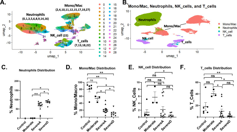
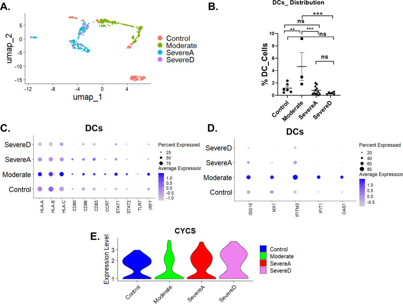
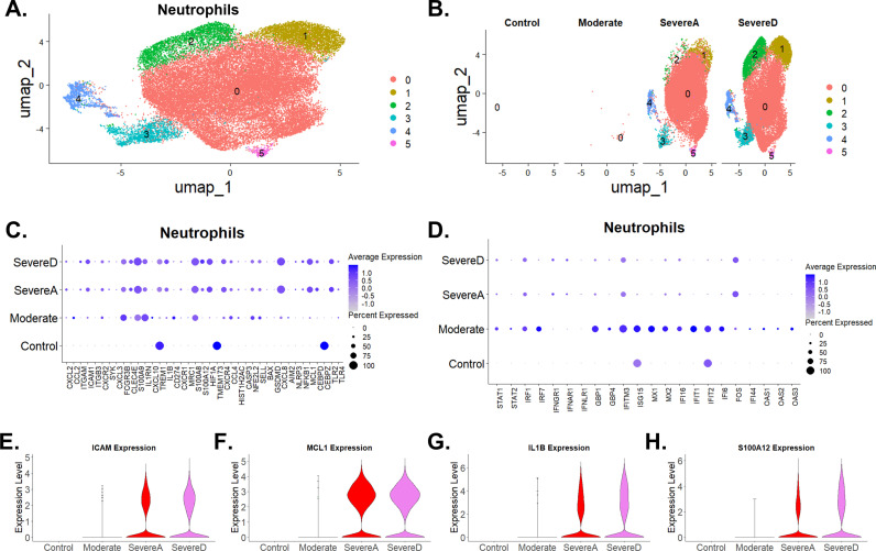
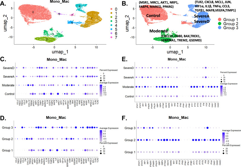
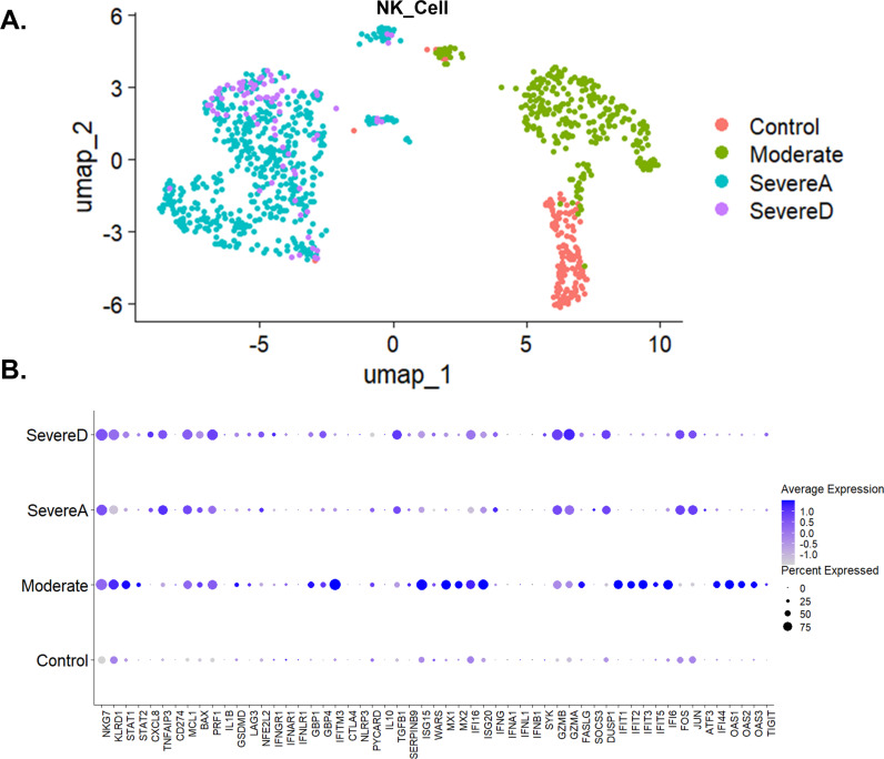
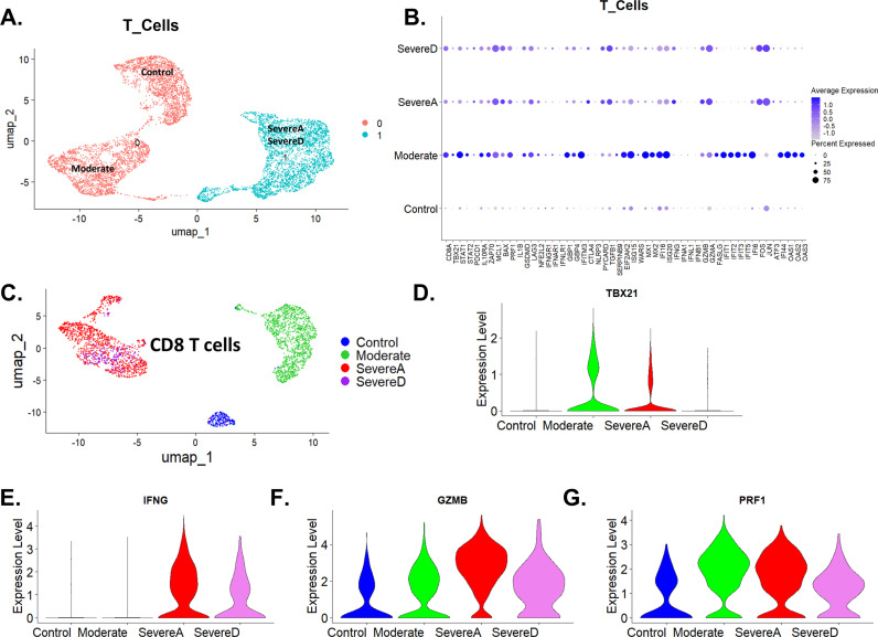
The continuous threats posed by Severe Acute Respiratory Syndrome Coronavirus 2 (SARS-CoV-2), the virus that causes COVID-19, including the emergence of potentially more infectious and deadly variants, necessitate ongoing studies to uncover novel and detailed mechanisms driving disease severity. Using single-cell transcriptomics, we conducted a secondary data analysis of bronchoalveolar lavage fluid (BALF) from COVID-19 patients of varying severities and healthy controls to comprehensively examine immune responses. We observed significant immune cell alterations correlating with disease severity. In severe cases, macrophages showed upregulation of pro-inflammatory genes TNFα and IL1β, contributing to severe inflammation and tissue damage. Neutrophils exhibited increased activation, marked by S100A8, CXCL8, and IL1β expression, with extended viability and reduced phagocytosis. Genes such as MCL1 and HIF1α supported extended viability, while MSR1 and MRC1 indicated reduced phagocytosis. Enhanced formation of neutrophil extracellular traps (NETs) and reduced clearance, indicated by NET-associated markers, were linked to thrombo-inflammation and organ damage. Both macrophages and neutrophils in severe cases showed impaired efferocytosis, indicated by decreased expression of MSR1 and TREM2 in macrophages and downregulation of FCGR3B in neutrophils, leading to the accumulation of apoptotic cells and exacerbating inflammation. Severe cases were characterized by M1 macrophages with high TNFα and IL1β, while milder cases had M2 macrophages with elevated PPARγ. Dendritic cells (DCs) in severe cases exhibited reduced proportions and attenuated expression of MHC class I genes (HLA-A, HLA-B, HLA-C) and co-stimulatory molecules (CD80, CD86), alongside increased cytochrome c expression, indicating impaired antigen presentation and enhanced apoptosis. NK and T cells in severe cases demonstrated altered receptor and gene expression, with increased activation markers IFNγ and ISG15, suggesting a paradoxical state of activation and exhaustion. This analysis highlights the critical role of dysregulated neutrophil, macrophage, dendritic cell, NK, and T cell responses in severe COVID-19, identifying potential therapeutic targets and providing novel insights into the disease.
严重急性呼吸综合征冠状病毒2(SARS-CoV-2)是导致新冠肺炎的病毒,它带来的持续威胁,包括可能出现更具传染性和致命性的变种,使得有必要持续开展研究以揭示驱动疾病严重程度的新的详细机制。我们利用单细胞转录组学技术,对不同严重程度的新冠肺炎患者和健康对照者的支气管肺泡灌洗液(BALF)进行了二次数据分析,以全面检查免疫反应。我们观察到与疾病严重程度相关的显著免疫细胞改变。在重症病例中,巨噬细胞表现出促炎基因TNFα和IL1β的上调,导致严重炎症和组织损伤。中性粒细胞表现出激活增加,以S100A8、CXCL8和IL1β的表达为标志,其存活期延长且吞噬作用降低。诸如MCL1和HIF1α等基因支持存活期延长,而MSR1和MRC1则表明吞噬作用降低。中性粒细胞胞外陷阱(NETs)形成增强且清除减少,这由NET相关标志物表明,与血栓炎症和器官损伤有关。重症病例中的巨噬细胞和中性粒细胞均表现出吞噬作用受损,这由巨噬细胞中MSR1和TREM2表达降低以及中性粒细胞中FCGR3B下调表明,导致凋亡细胞积累并加剧炎症。重症病例的特征是具有高TNFα和IL1β的M1巨噬细胞,而轻症病例则具有PPARγ升高的M2巨噬细胞。重症病例中的树突状细胞(DCs)比例降低,MHC I类基因(HLA-A、HLA-B、HLA-C)和共刺激分子(CD80、CD86)的表达减弱,同时细胞色素c表达增加,表明抗原呈递受损且凋亡增强。重症病例中的自然杀伤细胞(NK)和T细胞表现出受体和基因表达改变,激活标志物IFNγ和ISG15增加,表明存在激活和耗竭的矛盾状态。该分析突出了中性粒细胞、巨噬细胞、树突状细胞、NK细胞和T细胞反应失调在重症新冠肺炎中的关键作用,确定了潜在的治疗靶点,并为该疾病提供了新的见解。