• 文献检索
  • 文档翻译
  • 深度研究
  • 学术资讯
  • Suppr Zotero 插件Zotero 插件
  • 邀请有礼
  • 套餐&价格
  • 历史记录
应用&插件
Suppr Zotero 插件Zotero 插件浏览器插件Mac 客户端Windows 客户端微信小程序
定价
高级版会员购买积分包购买API积分包
服务
文献检索文档翻译深度研究API 文档MCP 服务
关于我们
关于 Suppr公司介绍联系我们用户协议隐私条款
关注我们

Suppr 超能文献

核心技术专利:CN118964589B侵权必究
粤ICP备2023148730 号-1Suppr @ 2026

文献检索

告别复杂PubMed语法,用中文像聊天一样搜索,搜遍4000万医学文献。AI智能推荐,让科研检索更轻松。

立即免费搜索

文件翻译

保留排版,准确专业,支持PDF/Word/PPT等文件格式,支持 12+语言互译。

免费翻译文档

深度研究

AI帮你快速写综述,25分钟生成高质量综述,智能提取关键信息,辅助科研写作。

立即免费体验

一种表示桡骨远端关节面三维倾斜度的新方法。

A Novel Method to Represent the Three-Dimensional Inclination of the Distal Radius Joint Surface.

作者信息

Ikumi Akira, Asai Reo, Eda Yusuke, Uchida Tooru, Kohyama Sho, Ogawa Takeshi, Yoshii Yuichi

机构信息

Department of Orthopedic Surgery, Institute of Medicine, University of Tsukuba, Tsukuba 305-8577, Ibaraki, Japan.

Department of Orthopedic Surgery, Tsukuba Medical Center Hospital, Tsukuba 305-8558, Ibaraki, Japan.

出版信息

Diagnostics (Basel). 2025 Feb 1;15(3):345. doi: 10.3390/diagnostics15030345.

DOI:10.3390/diagnostics15030345
PMID:39941275
原文链接:https://pmc.ncbi.nlm.nih.gov/articles/PMC11817736/
Abstract

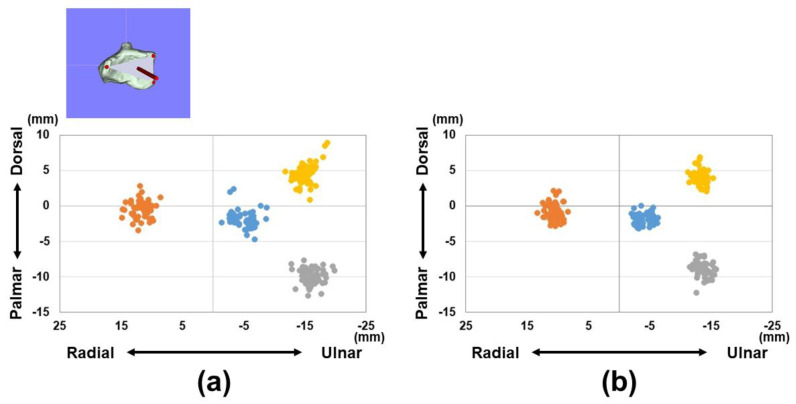
This study aims to define three-dimensional (3D) parameters for the inclination of the distal radius joint surface. The goal is to develop standardized parameters for fracture reduction through comprehensive 3D evaluations of the joint surfaces. We analyzed 112 CT scans of unaffected wrists (56 males and 56 females) to construct 3D models of the distal radius. Using 3D coordinates, the normal vectors and angles were calculated based on three reference points on the distal radius joint surface. These normal vector components were then converted into unit vector components A, B, and C for the x, y, and z axes, respectively. Additionally, the angles of these unit vectors were assessed in the xy, yz, and xz planes. The 3D measurements were compared between males and females and against traditional two-dimensional (2D) parameters such as palmar tilt and radial inclination. For males, the unit vector components were as follows: A: -0.14 ± 0.09, B: -0.92 ± 0.02, and C: -0.36 ± 0.07; for females, A: -0.21 ± 0.08, B: -0.90 ± 0.03, and C: -0.36 ± 0.05. Significant differences were found between males and females for the A and B vector components (representing the palmar-dorsal and proximal-distal axes, < 0.01). The angles of the unit vectors in the xy, yz, and xz planes were 8.9 ± 5.4°/12.9 ± 5.0°, 21.3 ± 4.1°/22.1 ± 3.2°, and 22.2 ± 14.8°/28.8 ± 10.1° for males and females, respectively. There were significant differences between males and females in the angles of the xy and xz planes (sagittal and axial planes, < 0.01). Strong correlations were observed between the xy-plane vectors and palmar tilt (r = 0.96), as well as between the yz-plane vectors and radial inclination (r = 0.88). This study evaluated the 3D inclination of the distal radius joint surface, revealing significant gender differences. This method, which also allows for the assessment of rotational alignment-difficult with conventional techniques-is expected to be a key 3D parameter in treating distal radius fractures.

摘要

本研究旨在确定桡骨远端关节面倾斜度的三维(3D)参数。目标是通过对关节面进行全面的3D评估来制定骨折复位的标准化参数。我们分析了112例未受影响手腕的CT扫描(56例男性和56例女性),以构建桡骨远端的3D模型。利用3D坐标,基于桡骨远端关节面上的三个参考点计算法向量和角度。然后将这些法向量分量分别转换为x、y和z轴的单位向量分量A、B和C。此外,在xy、yz和xz平面中评估这些单位向量的角度。比较了男性和女性之间的3D测量结果,并与传统的二维(2D)参数(如掌倾角和桡骨倾斜度)进行了对比。对于男性,单位向量分量如下:A:-0.14±0.09,B:-0.92±0.02,C:-0.36±0.07;对于女性,A:-0.21±0.08,B:-0.90±0.03,C:-0.36±0.05。在A和B向量分量(分别代表掌背和近远轴)方面,男性和女性之间存在显著差异(<0.01)。男性和女性在xy、yz和xz平面中单位向量的角度分别为8.9±5.4°/12.9±5.0°、21.3±4.1°/22.1±3.2°和22.2±14.8°/28.8±10.1°。男性和女性在xy和xz平面(矢状面和轴面)的角度存在显著差异(<0.01)。观察到xy平面向量与掌倾角之间存在强相关性(r = 0.96),以及yz平面向量与桡骨倾斜度之间存在强相关性(r = 0.88)。本研究评估了桡骨远端关节面的3D倾斜度,揭示了显著的性别差异。这种方法还能够评估旋转对线情况——这是传统技术难以做到的——有望成为治疗桡骨远端骨折的关键3D参数。

https://cdn.ncbi.nlm.nih.gov/pmc/blobs/056a/11817736/37dd3f232c36/diagnostics-15-00345-g007.jpg
https://cdn.ncbi.nlm.nih.gov/pmc/blobs/056a/11817736/fd470d24dfa0/diagnostics-15-00345-g001.jpg
https://cdn.ncbi.nlm.nih.gov/pmc/blobs/056a/11817736/67f6cf0f6964/diagnostics-15-00345-g002.jpg
https://cdn.ncbi.nlm.nih.gov/pmc/blobs/056a/11817736/99ab431b8a03/diagnostics-15-00345-g003.jpg
https://cdn.ncbi.nlm.nih.gov/pmc/blobs/056a/11817736/b492649c1fdc/diagnostics-15-00345-g004.jpg
https://cdn.ncbi.nlm.nih.gov/pmc/blobs/056a/11817736/fb6f89c2fbe9/diagnostics-15-00345-g005.jpg
https://cdn.ncbi.nlm.nih.gov/pmc/blobs/056a/11817736/4f03990983b8/diagnostics-15-00345-g006.jpg
https://cdn.ncbi.nlm.nih.gov/pmc/blobs/056a/11817736/37dd3f232c36/diagnostics-15-00345-g007.jpg

相似文献

1
A Novel Method to Represent the Three-Dimensional Inclination of the Distal Radius Joint Surface.一种表示桡骨远端关节面三维倾斜度的新方法。
Diagnostics (Basel). 2025 Feb 1;15(3):345. doi: 10.3390/diagnostics15030345.
2
A novel approach for three-dimensional evaluation of reduction morphology in distal radius fracture.一种用于评估桡骨远端骨折复位形态的三维新方法。
BMC Musculoskelet Disord. 2025 Feb 15;26(1):156. doi: 10.1186/s12891-025-08367-8.
3
Computer-Aided Assessment of Three-Dimensional Standard Bone Morphology of the Distal Radius.计算机辅助评估桡骨远端的三维标准骨形态
Diagnostics (Basel). 2022 Dec 17;12(12):3212. doi: 10.3390/diagnostics12123212.
4
Three-Dimensional Versus Radiographic Measurements for Analyzing Extra-Articular Distal Radius Malunion.三维测量与影像学测量在分析关节外桡骨远端畸形愈合中的应用对比。
J Hand Surg Am. 2020 Oct;45(10):984.e1-984.e7. doi: 10.1016/j.jhsa.2020.03.009. Epub 2020 Apr 20.
5
Three-dimensional evaluations of preoperative planning reproducibility for the osteosynthesis of distal radius fractures.桡骨远端骨折内固定术前规划可重复性的三维评估。
J Orthop Surg Res. 2021 Feb 12;16(1):131. doi: 10.1186/s13018-021-02278-9.
6
Three-Dimensional Morphometric Analysis of the Volar Cortical Shape of the Lunate Facet of the Distal Radius.桡骨远端月骨小关节掌侧皮质形态的三维形态计量学分析
Diagnostics (Basel). 2024 Aug 18;14(16):1802. doi: 10.3390/diagnostics14161802.
7
[Preliminary application of human-computer interaction CT imaging AI recognition and positioning technology in the treatment of type C1 distal radius fractures].人机交互CT成像人工智能识别与定位技术在C1型桡骨远端骨折治疗中的初步应用
Zhongguo Gu Shang. 2025 Jan 25;38(1):31-40. doi: 10.12200/j.issn.1003-0034.20240601.
8
Is a Novel Fluoroscopic Intraoperative Reference System Superior to Conventional Management for Distal Radius Fracture Reduction? A Propensity-matched Comparative Study.新型术中透视参考系统在桡骨远端骨折复位中优于常规管理吗?一项倾向评分匹配的对比研究。
Clin Orthop Relat Res. 2024 Mar 1;482(3):526-533. doi: 10.1097/CORR.0000000000002829. Epub 2023 Sep 6.
9
An Evaluation of the Reliability of Manual Landmark Identification on 3D Segmented Wrists.对手腕 3D 分割后手动标志点识别可靠性的评估。
J Bone Joint Surg Am. 2024 Feb 21;106(4):315-322. doi: 10.2106/JBJS.23.00173. Epub 2023 Nov 23.
10
Image-Based Comparison Between the Bilateral Symmetry of the Distal Radii Through Established Measures.通过既定测量方法对双侧桡骨远端对称性进行基于图像的比较。
J Hand Surg Am. 2019 Nov;44(11):966-972. doi: 10.1016/j.jhsa.2019.05.021. Epub 2019 Jul 13.

本文引用的文献

1
Relationship between Physical Characteristics and Morphological Features of the Articular Radius Surface: A Retrospective Single-Center Study.桡骨关节面物理特征与形态学特征的关系:一项回顾性单中心研究
Diagnostics (Basel). 2024 Sep 10;14(18):2005. doi: 10.3390/diagnostics14182005.
2
Three-Dimensional Morphometric Analysis of the Volar Cortical Shape of the Lunate Facet of the Distal Radius.桡骨远端月骨小关节掌侧皮质形态的三维形态计量学分析
Diagnostics (Basel). 2024 Aug 18;14(16):1802. doi: 10.3390/diagnostics14161802.
3
Radiographic Morphometric Analysis of the Distal Radius in the Tamil Nadu Population: A Retrospective Study.
泰米尔纳德邦人群桡骨远端的影像学形态计量分析:一项回顾性研究
Cureus. 2024 Jun 12;16(6):e62226. doi: 10.7759/cureus.62226. eCollection 2024 Jun.
4
Traditional radiography versus computed tomography to assess reduced distal radius fractures.传统放射摄影与计算机断层扫描评估桡骨远端骨折。
Eur J Trauma Emerg Surg. 2024 Oct;50(5):2313-2321. doi: 10.1007/s00068-024-02598-5. Epub 2024 Jul 10.
5
3D-assisted corrective osteotomies of the distal radius: a comparison of pre-contoured conventional implants versus patient-specific implants.3D 辅助下桡骨远端矫正截骨术:预制常规植入物与患者特异性植入物的比较。
Eur J Trauma Emerg Surg. 2024 Feb;50(1):37-47. doi: 10.1007/s00068-023-02415-5. Epub 2024 Jan 23.
6
A comparison of 3-D CT and 2-D plain radiograph measurements of the wrist in extra-articular malunited fractures of the distal radius.桡骨远端关节外骨折畸形愈合时腕关节的三维CT与二维平片测量比较
J Hand Surg Eur Vol. 2024 May;49(5):546-553. doi: 10.1177/17531934231213790. Epub 2023 Nov 21.
7
A three-dimensional computed tomography study of the palmar ulnar corner fragment in distal radial fractures.掌侧尺侧角碎片在桡骨远端骨折中的三维 CT 研究。
J Hand Surg Eur Vol. 2024 Mar;49(3):300-309. doi: 10.1177/17531934231211570. Epub 2023 Nov 16.
8
Computer-Aided Assessment of Three-Dimensional Standard Bone Morphology of the Distal Radius.计算机辅助评估桡骨远端的三维标准骨形态
Diagnostics (Basel). 2022 Dec 17;12(12):3212. doi: 10.3390/diagnostics12123212.
9
A Two-Step Approach for 3D-Guided Patient-Specific Corrective Limb Osteotomies.一种用于三维引导的患者特异性肢体矫正截骨术的两步法。
J Pers Med. 2022 Sep 6;12(9):1458. doi: 10.3390/jpm12091458.
10
Load Distribution in Dorsally-Angulated Distal Radius Deformity Using Finite Element Analysis.使用有限元分析评估背侧成角型桡骨远端畸形中的负荷分布
J Hand Surg Am. 2023 Oct;48(10):1062.e1-1062.e6. doi: 10.1016/j.jhsa.2022.02.022. Epub 2022 Aug 13.