• 文献检索
  • 文档翻译
  • 深度研究
  • 学术资讯
  • Suppr Zotero 插件Zotero 插件
  • 邀请有礼
  • 套餐&价格
  • 历史记录
应用&插件
Suppr Zotero 插件Zotero 插件浏览器插件Mac 客户端Windows 客户端微信小程序
定价
高级版会员购买积分包购买API积分包
服务
文献检索文档翻译深度研究API 文档MCP 服务
关于我们
关于 Suppr公司介绍联系我们用户协议隐私条款
关注我们

Suppr 超能文献

核心技术专利:CN118964589B侵权必究
粤ICP备2023148730 号-1Suppr @ 2026

文献检索

告别复杂PubMed语法,用中文像聊天一样搜索,搜遍4000万医学文献。AI智能推荐,让科研检索更轻松。

立即免费搜索

文件翻译

保留排版,准确专业,支持PDF/Word/PPT等文件格式,支持 12+语言互译。

免费翻译文档

深度研究

AI帮你快速写综述,25分钟生成高质量综述,智能提取关键信息,辅助科研写作。

立即免费体验

胎儿发育过程中人类胸腺微环境的形态学特征。

Morphological characteristics of microenvironment in the human thymus during fetal development.

作者信息

Maletin Nemanja, Denda Nikola, Borocki Stefan, Golušin Zoran, Rašković Aleksandar, Fejsa-Levakov Aleksandra, Višnjić Bojana Andrejić, Amidžić Jelena

机构信息

Faculty of Medicine, University of Novi Sad, Novi Sad, Serbia.

出版信息

BMC Res Notes. 2025 Mar 3;18(1):92. doi: 10.1186/s13104-025-07109-2.

DOI:10.1186/s13104-025-07109-2
PMID:40033348
原文链接:https://pmc.ncbi.nlm.nih.gov/articles/PMC11877800/
Abstract

BACKGROUND

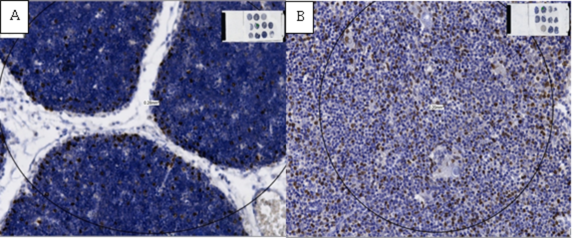
The thymus is a key organ for the development of T cells. T cell precursors first migrate from the bone marrow to the thymus. During maturation, these precursors require interactions with various types of cells that form the thymic microenvironment, such as epithelial, mesenchymal, and other immune cells not belonging to the T lineage. The aim of this study was to examine the changes in the number and diameter of Hassall's corpuscles, as well as the density and distribution of epithelial cells (p63+) and macrophages (CD68+).

METHODS

Twenty-five fetal thymus samples were examined, divided into five groups according to gestational age. The samples were processed using standard histological methods and immunohistochemical staining.

RESULTS

The study showed that the number and diameter of Hassall's corpuscles gradually increased during fetal development, with a significant increase from the 14th to the 38th gestational week. The average diameter of Hassall's corpuscles was largest in the age group of 34-38 weeks. The density of p63 + epithelial cells decreased in correlation with gestational week, while the density of CD68 + macrophages significantly increased, particularly in the thymic medulla, towards the end of the fetal period.

CONCLUSIONS

An increase in the number and size of Hassall's corpuscles during fetal development was recorded, while the density of epithelial cells decreased and the density of macrophages increased.

摘要

背景

胸腺是T细胞发育的关键器官。T细胞前体首先从骨髓迁移至胸腺。在成熟过程中,这些前体需要与构成胸腺微环境的各种类型细胞相互作用,如上皮细胞、间充质细胞以及其他不属于T谱系的免疫细胞。本研究的目的是检测哈氏小体的数量和直径变化,以及上皮细胞(p63+)和巨噬细胞(CD68+)的密度和分布。

方法

检查了25份胎儿胸腺样本,根据胎龄分为五组。样本采用标准组织学方法和免疫组织化学染色进行处理。

结果

研究表明,哈氏小体的数量和直径在胎儿发育过程中逐渐增加,从妊娠第14周到第38周有显著增加。哈氏小体的平均直径在34 - 38周龄组最大。p63+上皮细胞的密度随孕周增加而降低,而CD68+巨噬细胞的密度显著增加,尤其是在胎儿期末期的胸腺髓质中。

结论

记录到胎儿发育过程中哈氏小体的数量和大小增加,而上皮细胞密度降低,巨噬细胞密度增加。

https://cdn.ncbi.nlm.nih.gov/pmc/blobs/d99f/11877800/50daae610659/13104_2025_7109_Fig9_HTML.jpg
https://cdn.ncbi.nlm.nih.gov/pmc/blobs/d99f/11877800/699de6384bcb/13104_2025_7109_Fig1_HTML.jpg
https://cdn.ncbi.nlm.nih.gov/pmc/blobs/d99f/11877800/25975a17da3b/13104_2025_7109_Fig2_HTML.jpg
https://cdn.ncbi.nlm.nih.gov/pmc/blobs/d99f/11877800/d959719fbab2/13104_2025_7109_Fig3_HTML.jpg
https://cdn.ncbi.nlm.nih.gov/pmc/blobs/d99f/11877800/8e3aa6d2c8ee/13104_2025_7109_Fig4_HTML.jpg
https://cdn.ncbi.nlm.nih.gov/pmc/blobs/d99f/11877800/4e5e00241414/13104_2025_7109_Fig5_HTML.jpg
https://cdn.ncbi.nlm.nih.gov/pmc/blobs/d99f/11877800/f7907b6a0158/13104_2025_7109_Fig6_HTML.jpg
https://cdn.ncbi.nlm.nih.gov/pmc/blobs/d99f/11877800/ffafb39ddc77/13104_2025_7109_Fig7_HTML.jpg
https://cdn.ncbi.nlm.nih.gov/pmc/blobs/d99f/11877800/0a0f63e5a5bc/13104_2025_7109_Fig8_HTML.jpg
https://cdn.ncbi.nlm.nih.gov/pmc/blobs/d99f/11877800/50daae610659/13104_2025_7109_Fig9_HTML.jpg

相似文献

1
Morphological characteristics of microenvironment in the human thymus during fetal development.胎儿发育过程中人类胸腺微环境的形态学特征。
BMC Res Notes. 2025 Mar 3;18(1):92. doi: 10.1186/s13104-025-07109-2.
2
What do we know about the structure of human thymic Hassall's corpuscles? A histochemical, immunohistochemical, and electron microscopic study.我们对人类胸腺哈氏小体的结构了解多少?一项组织化学、免疫组织化学和电子显微镜研究。
Ann Anat. 2017 May;211:140-148. doi: 10.1016/j.aanat.2017.02.006. Epub 2017 Mar 6.
3
Hassall's corpuscles instruct dendritic cells to induce CD4+CD25+ regulatory T cells in human thymus.哈氏小体指导树突状细胞在人类胸腺中诱导产生CD4+CD25+调节性T细胞。
Nature. 2005 Aug 25;436(7054):1181-5. doi: 10.1038/nature03886.
4
The normal human newborns thymus.正常人类新生儿的胸腺。
Bratisl Lek Listy. 2013;114(7):402-8. doi: 10.4149/bll_2013_086.
5
Immunohistochemical study of tyrosine phosphorylation signaling in Hassall's corpuscles of the human thymus.人胸腺哈氏小体中酪氨酸磷酸化信号的免疫组织化学研究。
Acta Histochem. 1999 Nov;101(4):421-9. doi: 10.1016/s0065-1281(99)80042-7.
6
TGF-β type II receptor expression in thymic epithelial cells inhibits the development of Hassall's corpuscles in mice.TGF-β 型 II 受体在胸腺上皮细胞中的表达抑制了小鼠 Hassall 小体的发育。
Int Immunol. 2013 Nov;25(11):633-42. doi: 10.1093/intimm/dxt026. Epub 2013 Aug 8.
7
Epithelial cell heterogeneity in mammalian thymus: monoclonal antibody to high molecular weight keratins exclusively binds to Hassall's corpuscles.哺乳动物胸腺中的上皮细胞异质性:针对高分子量角蛋白的单克隆抗体仅与哈氏小体结合。
Histochem J. 1989 Jun;21(6):357-64. doi: 10.1007/BF01798499.
8
[Morphology of Hassall's corpuscles and their microenvironment in neonates with increased thymus weight].[胸腺重量增加的新生儿中哈氏小体的形态及其微环境]
Arkh Patol. 2024;86(1):13-20. doi: 10.17116/patol20248601113.
9
The first histological and immunohistochemical examination of thymus in a case of fetus in fetu.胎儿内生儿一例的胸腺组织学和免疫组织化学检查。
Ann Anat. 2010 Aug 20;192(4):232-6. doi: 10.1016/j.aanat.2010.05.007. Epub 2010 Jun 11.
10
The human thymic microenvironment: thymic epithelium contains specific keratins associated with early and late stages of epidermal keratinocyte maturation.人类胸腺微环境:胸腺上皮含有与表皮角质形成细胞成熟早期和晚期相关的特定角蛋白。
Differentiation. 1986;31(1):67-77. doi: 10.1111/j.1432-0436.1986.tb00385.x.

本文引用的文献

1
The initial age-associated decline in early T-cell progenitors reflects fewer pre-thymic progenitors and altered signals in the bone marrow and thymus microenvironments.早期 T 细胞前体细胞随年龄的初始下降反映了骨髓和胸腺微环境中前胸腺祖细胞数量减少和信号改变。
Aging Cell. 2023 Aug;22(8):e13870. doi: 10.1111/acel.13870. Epub 2023 May 23.
2
Thymic Microenvironment: Interactions Between Innate Immune Cells and Developing Thymocytes.胸腺微环境:固有免疫细胞与发育中的胸腺细胞的相互作用。
Front Immunol. 2022 Apr 8;13:885280. doi: 10.3389/fimmu.2022.885280. eCollection 2022.
3
Ephrin Receptors (Ephs) Expression in Thymic Epithelial Tumors: Prognostic Implications and Future Therapeutic Approaches.
胸腺上皮肿瘤中 Ephrin 受体(Ephs)的表达:预后意义及未来治疗方法
Diagnostics (Basel). 2021 Dec 3;11(12):2265. doi: 10.3390/diagnostics11122265.
4
The thymus: General concepts on embryology, anatomy, histology and immunohistochemistry.胸腺:胚胎学、解剖学、组织学和免疫组织化学的一般概念。
Semin Diagn Pathol. 2022 Mar;39(2):86-91. doi: 10.1053/j.semdp.2021.06.003. Epub 2021 Jun 10.
5
Thymus-Pineal Gland Axis: Revisiting Its Role in Human Life and Ageing.胸腺-松果腺轴:重新探讨其在人类生命和衰老中的作用。
Int J Mol Sci. 2020 Nov 20;21(22):8806. doi: 10.3390/ijms21228806.
6
Implications of Oxidative Stress and Cellular Senescence in Age-Related Thymus Involution.氧化应激和细胞衰老对与年龄相关的胸腺萎缩的影响。
Oxid Med Cell Longev. 2020 Feb 5;2020:7986071. doi: 10.1155/2020/7986071. eCollection 2020.
7
Thymic Epithelial Cells Contribute to Thymopoiesis and T Cell Development.胸腺上皮细胞有助于胸腺生成和 T 细胞发育。
Front Immunol. 2020 Jan 31;10:3099. doi: 10.3389/fimmu.2019.03099. eCollection 2019.
8
Hassall's corpuscles with cellular-senescence features maintain IFNα production through neutrophils and pDC activation in the thymus.具有细胞衰老特征的 Hassall 小体通过中性粒细胞和 pDC 激活维持胸腺中 IFNα 的产生。
Int Immunol. 2019 Mar 5;31(3):127-139. doi: 10.1093/intimm/dxy073.
9
Macrophage Clearance of Apoptotic Cells: A Critical Assessment.巨噬细胞清除凋亡细胞:一项关键性评估。
Front Immunol. 2018 Jan 30;9:127. doi: 10.3389/fimmu.2018.00127. eCollection 2018.
10
What do we know about the structure of human thymic Hassall's corpuscles? A histochemical, immunohistochemical, and electron microscopic study.我们对人类胸腺哈氏小体的结构了解多少?一项组织化学、免疫组织化学和电子显微镜研究。
Ann Anat. 2017 May;211:140-148. doi: 10.1016/j.aanat.2017.02.006. Epub 2017 Mar 6.