• 文献检索
  • 文档翻译
  • 深度研究
  • 学术资讯
  • Suppr Zotero 插件Zotero 插件
  • 邀请有礼
  • 套餐&价格
  • 历史记录
应用&插件
Suppr Zotero 插件Zotero 插件浏览器插件Mac 客户端Windows 客户端微信小程序
定价
高级版会员购买积分包购买API积分包
服务
文献检索文档翻译深度研究API 文档MCP 服务
关于我们
关于 Suppr公司介绍联系我们用户协议隐私条款
关注我们

Suppr 超能文献

核心技术专利:CN118964589B侵权必究
粤ICP备2023148730 号-1Suppr @ 2026

文献检索

告别复杂PubMed语法,用中文像聊天一样搜索,搜遍4000万医学文献。AI智能推荐,让科研检索更轻松。

立即免费搜索

文件翻译

保留排版,准确专业,支持PDF/Word/PPT等文件格式,支持 12+语言互译。

免费翻译文档

深度研究

AI帮你快速写综述,25分钟生成高质量综述,智能提取关键信息,辅助科研写作。

立即免费体验

计算机断层扫描检测到的中颅窝副蝶骨孔。

An accessory sphenoidal foramen of the middle cranial fossa detected on computed tomography.

作者信息

Triantafyllou George, Papadopoulos-Manolarakis Panagiotis, Olewnik Łukasz, Duparc Fabrice, Tsakotos George, Zielinska Nicol, Piagkou Maria

机构信息

Department of Anatomy, School of Medicine, Faculty of Health Sciences, National and Kapodistrian University of Athens, 75 Mikras Asias Str., Goudi, 11527, Athens, Greece.

Department of Neurosurgery, General Hospital of Nikaia-Piraeus, Athens, Greece.

出版信息

Surg Radiol Anat. 2025 Mar 10;47(1):89. doi: 10.1007/s00276-025-03601-3.

DOI:10.1007/s00276-025-03601-3
PMID:40063286
原文链接:https://pmc.ncbi.nlm.nih.gov/articles/PMC11893693/
Abstract

BACKGROUND

The skull base depicts significant morphological variability, which is frequently described due to its neurosurgical significance. The middle cranial fossa's accessory foramen has rarely been described.

MATERIALS

A 53-year-old female patient's computed tomography (CT) scan was further investigated for its unusual morphology.

RESULTS

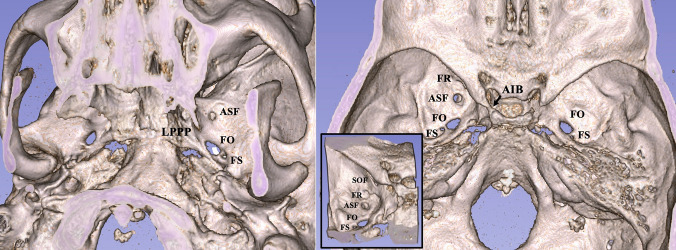
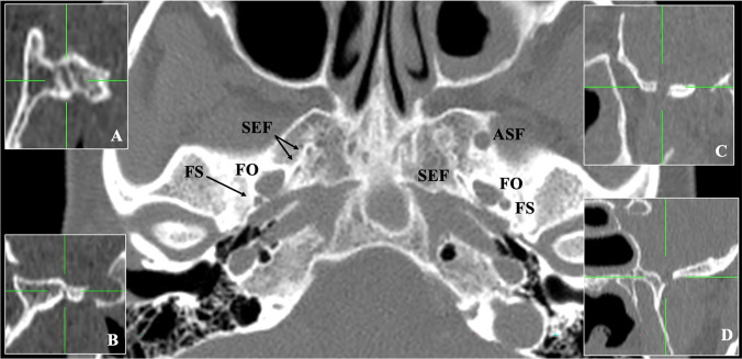
On the left-sided middle cranial fossa, an accessory sphenoidal foramen (ASF) was observed, located 3.3 mm posterior to the foramen rotundum (FR) and 5.5 mm anterior to the foramen ovale (FO). Extracranially, the ASF opened into the infratemporal fossa and coexisted with another sphenoidal emissary foramen (SEF), anteromedially to the FO. On the right side, two SEF were located anteromedially to the FO.

CONCLUSIONS

Similar to the current case, ASF of the middle cranial fossa were reported in a previous study with a prevalence of 0.20%. The unconstraint well described accessory foramina are the emissary foramina that transmit emissary veins, and are of interest for anatomists, radiologists and neurosurgeons.

摘要

背景

颅底呈现出显著的形态学变异性,因其在神经外科手术中的重要意义而经常被描述。中颅窝的副孔很少被描述。

材料

对一名53岁女性患者的计算机断层扫描(CT)进行了进一步研究,因其形态异常。

结果

在左侧中颅窝观察到一个蝶骨副孔(ASF),位于圆孔(FR)后方3.3毫米处,卵圆孔(FO)前方5.5毫米处。颅外,ASF开口于颞下窝,并与另一个蝶骨导静脉孔(SEF)共存,位于FO的前内侧。在右侧,两个SEF位于FO的前内侧。

结论

与当前病例类似,先前的一项研究报道中颅窝ASF的发生率为0.20%。未受严格描述的副孔是传导导静脉的导静脉孔,对解剖学家、放射科医生和神经外科医生具有重要意义。

https://cdn.ncbi.nlm.nih.gov/pmc/blobs/92a5/11893693/76d93b7b0cd3/276_2025_3601_Fig2_HTML.jpg
https://cdn.ncbi.nlm.nih.gov/pmc/blobs/92a5/11893693/c897f08f65ce/276_2025_3601_Fig1_HTML.jpg
https://cdn.ncbi.nlm.nih.gov/pmc/blobs/92a5/11893693/76d93b7b0cd3/276_2025_3601_Fig2_HTML.jpg
https://cdn.ncbi.nlm.nih.gov/pmc/blobs/92a5/11893693/c897f08f65ce/276_2025_3601_Fig1_HTML.jpg
https://cdn.ncbi.nlm.nih.gov/pmc/blobs/92a5/11893693/76d93b7b0cd3/276_2025_3601_Fig2_HTML.jpg

相似文献

1
An accessory sphenoidal foramen of the middle cranial fossa detected on computed tomography.计算机断层扫描检测到的中颅窝副蝶骨孔。
Surg Radiol Anat. 2025 Mar 10;47(1):89. doi: 10.1007/s00276-025-03601-3.
2
Concomitant Sphenopterygoid Canal and Duplicated Foramen Rotundum Detected on Computed Tomography Scan.计算机断层扫描检测到蝶翼管与重复的圆孔并存。
J Craniofac Surg. 2025 Jun 1;36(4):1381-1382. doi: 10.1097/SCS.0000000000011118. Epub 2025 Feb 6.
3
The sphenopterygoid canal and pterygoid foramen.蝶筛骨管和翼突孔。
Surg Radiol Anat. 2024 May;46(5):645-648. doi: 10.1007/s00276-024-03353-6. Epub 2024 Apr 2.
4
The sphenoidal emissary foramen and the emissary vein: Anatomy and clinical relevance.蝶骨导静脉孔和导静脉:解剖学及临床意义。
Clin Anat. 2020 Jul;33(5):767-781. doi: 10.1002/ca.23504. Epub 2019 Nov 15.
5
Unusual foramen in the middle cranial fossae of adult black South African skull specimens.成年南非黑人颅骨标本中颅窝的异常孔。
Surg Radiol Anat. 2017 Jul;39(7):815-818. doi: 10.1007/s00276-016-1780-0. Epub 2016 Nov 11.
6
Foramen venosum: a clinicoanatomic insight into its occurrence and morphometry.静脉孔:对其出现及形态测量的临床解剖学见解。
Surg Radiol Anat. 2023 Apr;45(4):409-415. doi: 10.1007/s00276-023-03106-x. Epub 2023 Feb 22.
7
The size of the foramen ovale regarding to the presence and absence of the emissary sphenoidal foramen: is there any relationship between them?关于蝶导静脉孔的有无,卵圆孔的大小:它们之间存在关系吗?
Folia Morphol (Warsz). 2018;77(1):90-98. doi: 10.5603/FM.a2017.0068. Epub 2017 Jul 13.
8
Anatomic Study on Sphenoidal Emissary Foramen by Using Cone-Beam Computed Tomography.使用锥形束计算机断层扫描对蝶导静脉孔的解剖学研究。
J Craniofac Surg. 2018 Jul;29(5):e477-e480. doi: 10.1097/SCS.0000000000004492.
9
Accessory spine of the foramen ovale.卵圆孔副棘
Folia Morphol (Warsz). 2012 Nov;71(4):263-6.
10
Variant Bilateral Foramina of the Middle Cranial Fossa.颅中窝双侧变异孔
Cureus. 2022 Dec 27;14(12):e33014. doi: 10.7759/cureus.33014. eCollection 2022 Dec.

引用本文的文献

1
The Developing Human Sphenoid Bone: Linking Embryological Development to Adult Morphology.发育中的人类蝶骨:将胚胎发育与成人形态联系起来。
Biology (Basel). 2025 Aug 20;14(8):1090. doi: 10.3390/biology14081090.

本文引用的文献

1
The sphenopterygoid canal and pterygoid foramen.蝶筛骨管和翼突孔。
Surg Radiol Anat. 2024 May;46(5):645-648. doi: 10.1007/s00276-024-03353-6. Epub 2024 Apr 2.
2
Variant Bilateral Foramina of the Middle Cranial Fossa.颅中窝双侧变异孔
Cureus. 2022 Dec 27;14(12):e33014. doi: 10.7759/cureus.33014. eCollection 2022 Dec.
3
The sphenoidal emissary foramina prevalence: a meta-analysis of 6,369 subjects.蝶骨导静脉孔出现率的荟萃分析:6369 例样本研究
Surg Radiol Anat. 2023 Jan;45(1):43-53. doi: 10.1007/s00276-022-03051-1. Epub 2022 Dec 6.
4
A giant foramen of Vesalius: case report.巨大维萨里孔:病例报告
Anat Cell Biol. 2022 Sep 30;55(3):373-375. doi: 10.5115/acb.22.017. Epub 2022 Jun 13.
5
Characterization of the pterygomeningeal artery based on branching pattern and muscular distribution.基于分支模式和肌肉分布对翼状缝动脉进行描述。
Surg Radiol Anat. 2022 Apr;44(4):543-550. doi: 10.1007/s00276-022-02911-0. Epub 2022 Mar 4.
6
Duplication of the foramen rotundum: A rare case report.圆孔重复:一例罕见病例报告。
Morphologie. 2023 Mar;107(356):138-141. doi: 10.1016/j.morpho.2021.11.004. Epub 2022 Feb 28.
7
The size of the foramen ovale regarding to the presence and absence of the emissary sphenoidal foramen: is there any relationship between them?关于蝶导静脉孔的有无,卵圆孔的大小:它们之间存在关系吗?
Folia Morphol (Warsz). 2018;77(1):90-98. doi: 10.5603/FM.a2017.0068. Epub 2017 Jul 13.
8
The revised anatomy of the canals connecting the orbit with the cranial cavity.连接眼眶与颅腔的管道的修订解剖结构。
Orbit. 2017 Apr;36(2):110-117. doi: 10.1080/01676830.2017.1279662. Epub 2017 Mar 3.
9
Unusual foramen in the middle cranial fossae of adult black South African skull specimens.成年南非黑人颅骨标本中颅窝的异常孔。
Surg Radiol Anat. 2017 Jul;39(7):815-818. doi: 10.1007/s00276-016-1780-0. Epub 2016 Nov 11.
10
Doubled foramen rotundum and maxillary nerve fenestration.圆孔及上颌神经开窗加倍。
Surg Radiol Anat. 2011 Oct;33(8):723-6. doi: 10.1007/s00276-011-0810-1. Epub 2011 Apr 5.