Okabe Naohiko, Wei Xiaofei, Abumeri Farah, Batac Jonathan, Hovanesyan Mary, Dai Weiye, Azarapetian Srbui, Campagna Jesus, Pilati Nadia, Marasco Agostino, Alvaro Giuseppe, Gunthorpe Martin J, Varghese John, Cramer Steven C, Mody Istvan, Carmichael S Thomas
Department of Neurology, David Geffen School of Medicine, UCLA, Los Angeles, CA, 90095, USA.
Department of Neurosurgery, David Geffen School of Medicine, UCLA, Los Angeles, CA, 90095, USA.
Nat Commun. 2025 Mar 15;16(1):2556. doi: 10.1038/s41467-025-57860-0.
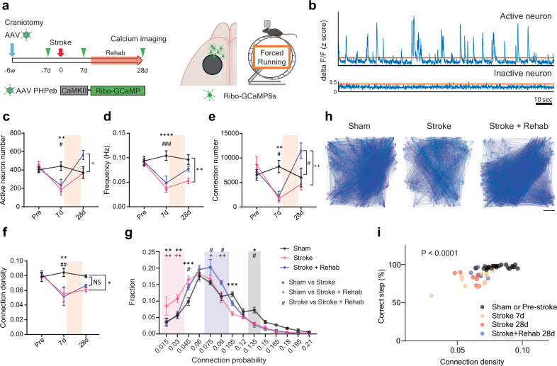
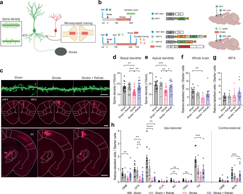
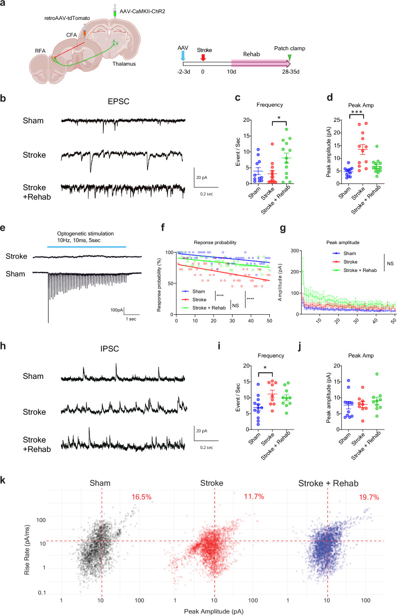
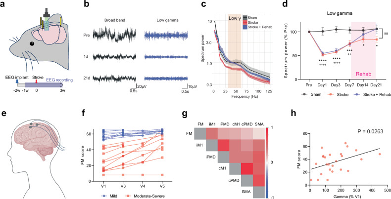
Motor disability is a critical impairment in stroke patients. Rehabilitation has a limited effect on recovery; but there is no medical therapy for post-stroke recovery. The biological mechanisms of rehabilitation in the brain remain unknown. Here, using a photothrombotic stroke model in male mice, we demonstrate that rehabilitation after stroke selectively enhances synapse formation in presynaptic parvalbumin interneurons and postsynaptic neurons in the rostral forelimb motor area with axonal projections to the caudal forelimb motor area where stroke was induced (stroke-projecting neuron). Rehabilitation improves motor performance and neuronal functional connectivity, while inhibition of stroke-projecting neurons diminishes motor recovery. Stroke-projecting neurons show decreased dendritic spine density, reduced external synaptic inputs, and a lower proportion of parvalbumin synapse in the total GABAergic input. Parvalbumin interneurons regulate neuronal functional connectivity, and their activation during training is necessary for recovery. Furthermore, gamma oscillation, a parvalbumin-regulated rhythm, is increased with rehabilitation-induced recovery in animals after stroke and stroke patients. Pharmacological enhancement of parvalbumin interneuron function improves motor recovery after stroke, reproducing rehabilitation recovery. These findings identify brain circuits that mediate rehabilitation-recovery and the possibility for rational selection of pharmacological agents to deliver the first molecular-rehabilitation therapeutic.
运动功能障碍是中风患者的一种关键损伤。康复治疗对恢复的效果有限;但目前尚无针对中风后恢复的药物治疗方法。大脑中康复的生物学机制仍不清楚。在此,我们利用雄性小鼠的光血栓性中风模型,证明中风后的康复治疗选择性地增强了前肢运动区前部的突触前小白蛋白中间神经元和突触后神经元中的突触形成,这些神经元的轴突投射到诱导中风的前肢运动区后部(中风投射神经元)。康复治疗改善了运动表现和神经元功能连接,而抑制中风投射神经元则会削弱运动恢复。中风投射神经元表现出树突棘密度降低、外部突触输入减少以及在总的γ-氨基丁酸能输入中,小白蛋白突触的比例较低。小白蛋白中间神经元调节神经元功能连接,并且它们在训练期间的激活对于恢复是必要的。此外,γ振荡是一种由小白蛋白调节的节律,在中风后的动物和中风患者中,随着康复诱导的恢复而增加。药理增强小白蛋白中间神经元功能可改善中风后的运动恢复,重现康复治疗的恢复效果。这些发现确定了介导康复恢复的脑回路,以及合理选择药物以提供首个分子康复治疗的可能性。