Zheng Henry W, Bui Alex A T, Ensrud Kristine E, Wright Nicole C, Manson JoAnn E, Watts Nelson B, Johnson Karen C, Shadyab Aladdin H, Crandall Carolyn J
Medical and Imaging Informatics, UCLA (University of California, Los Angeles), Los Angeles.
Departments of Radiological Sciences, Bioengineering and Bioinformatics, David Geffen School of Medicine at UCLA, Los Angeles.
JAMA Netw Open. 2025 Mar 3;8(3):e250626. doi: 10.1001/jamanetworkopen.2025.0626.
For younger postmenopausal women, clinical guidelines recommend using osteoporosis risk prediction tools to identify candidates with low bone mineral density (BMD). However, the performance of these tools is not well quantified.
To examine the performance of Osteoporosis Risk Assessment Instrument (ORAI) and Osteoporosis Index of Risk (OSIRIS), compared with Osteoporosis Self-Assessment Tool (OST), in identifying the presence of osteoporotic BMD in younger postmenopausal women.
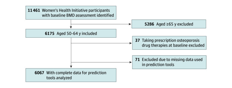
DESIGN, SETTING, AND PARTICIPANTS: This cross-sectional study used data from the Women's Health Initiative Bone Density Substudy, which was conducted at 3 clinical centers in Tucson and Phoenix, Arizona; Pittsburgh, Pennsylvania; and Birmingham, Alabama. Participants were healthy postmenopausal women aged 50 to 64 years with BMD measurements evaluated using the 3 risk prediction tools: OSIRIS, ORAI, and OST. Risk factors and other participant characteristics were compared across osteoporosis status. Data were collected from October 1993 to December 1998 and analyzed between September 23, 2023, and April 10, 2024.
The primary exposures were OSIRIS, ORAI, and OST risk scores.
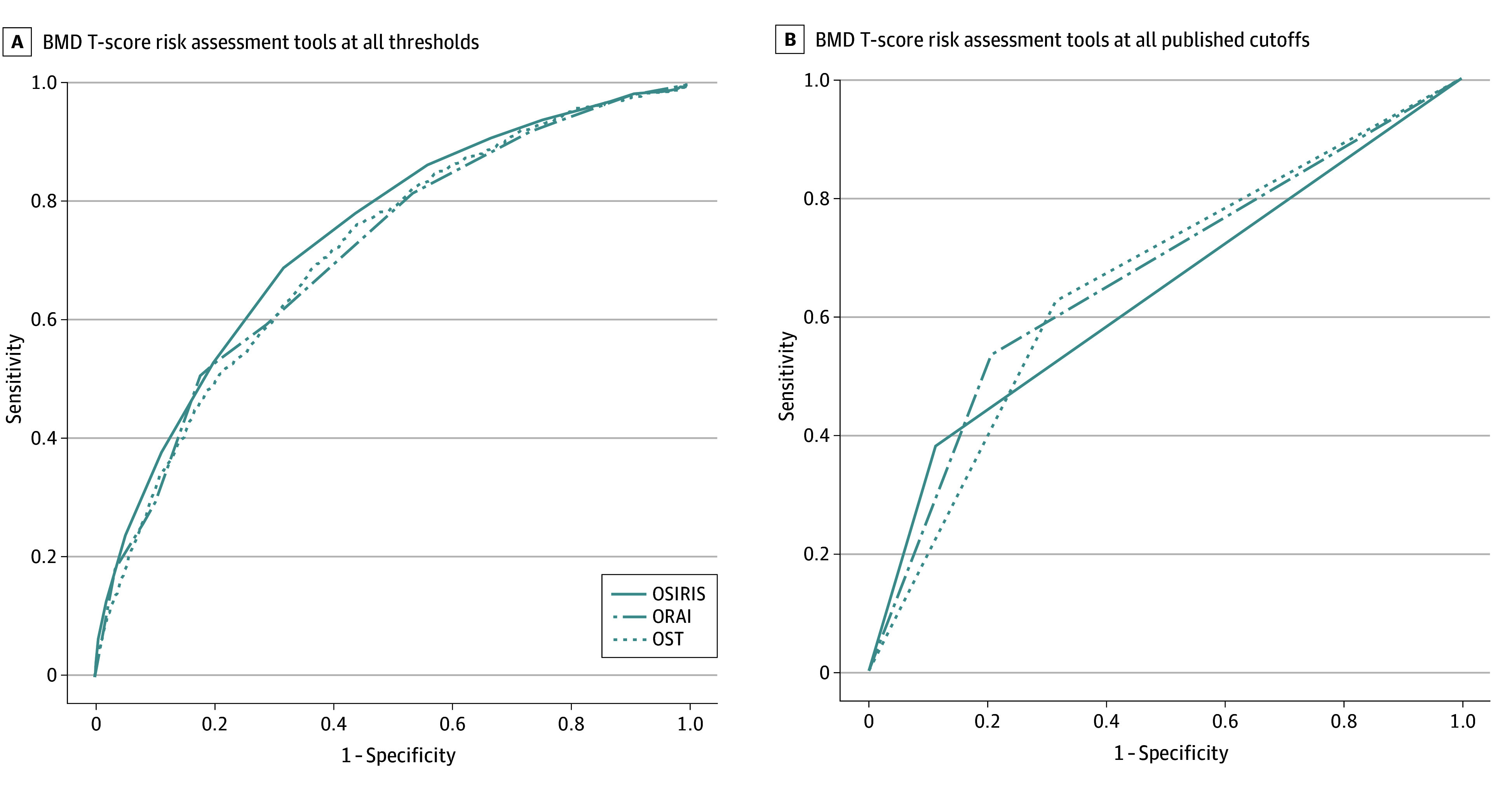
Primary outcome was osteoporosis defined by BMD T score of -2.5 or lower at 1 or more of 3 anatomical locations: femoral neck, total hip, and/or lumbar spine. The tools were evaluated via area under the receiver operating characteristic curve (AUC) at published score cutoffs and at alternate cutoffs.
Among 6067 included participants (mean [SD] age at baseline, 57.7 [4.1] years), the prevalence of osteoporosis was 14.1% (n = 857) at any 1 of 3 anatomical sites. AUC for identifying osteoporosis at any site was 0.633 (95% CI, 0.633-0.634) for OSIRIS, 0.663 (95% CI, 0.663-0.664) for ORAI, and 0.654 (95% CI, 0.654-0.655) for OST.
In this cross-sectional study, 3 guideline-recommended osteoporosis risk assessment tools had fair to moderate discrimination in identifying osteoporosis defined by lowest BMD at any 1 of 3 skeletal sites. Screening is essential to reducing individual and societal burden of osteoporosis and related fractures, and this study showed a gap in identifying younger postmenopausal women using common clinical risk factors.
对于绝经后较年轻女性,临床指南建议使用骨质疏松症风险预测工具来识别骨矿物质密度(BMD)较低的人群。然而,这些工具的性能尚未得到充分量化。
将骨质疏松症风险评估工具(ORAI)和骨质疏松症风险指数(OSIRIS)与骨质疏松症自我评估工具(OST)相比较,以检验它们在识别绝经后较年轻女性骨质疏松性BMD方面的性能。
设计、地点和参与者:这项横断面研究使用了来自女性健康倡议骨密度子研究的数据,该研究在亚利桑那州图森市和凤凰城、宾夕法尼亚州匹兹堡市以及阿拉巴马州伯明翰市的3个临床中心进行。参与者为年龄在50至64岁之间的健康绝经后女性,她们使用三种风险预测工具(OSIRIS、ORAI和OST)进行了BMD测量。比较了骨质疏松症状态下的风险因素和其他参与者特征。数据收集于1993年10月至1998年12月,并于2023年9月23日至2024年4月10日进行分析。
主要暴露因素为OSIRIS、ORAI和OST风险评分。
主要结局是骨质疏松症,定义为在股骨颈、全髋和/或腰椎这三个解剖部位中的至少一个部位,BMD T评分≤-2.5。通过在已公布的评分临界值和替代临界值下的受试者工作特征曲线下面积(AUC)对这些工具进行评估。
在纳入的6067名参与者中(基线时平均[标准差]年龄为57.7[4.1]岁),在三个解剖部位中的任何一个部位,骨质疏松症的患病率为14.1%(n = 857)。OSIRIS识别任何部位骨质疏松症的AUC为0.633(95%CI,0.633 - 0.634),ORAI为0.663(95%CI,0.663 - 0.664),OST为0.654(95%CI,0.654 - 0.655)。
在这项横断面研究中,三种指南推荐的骨质疏松症风险评估工具在识别由三个骨骼部位中任何一个部位的最低BMD定义的骨质疏松症方面,具有中等偏下的鉴别能力。筛查对于减轻骨质疏松症及相关骨折的个人和社会负担至关重要,但本研究表明,在使用常见临床风险因素识别绝经后较年轻女性方面存在差距。